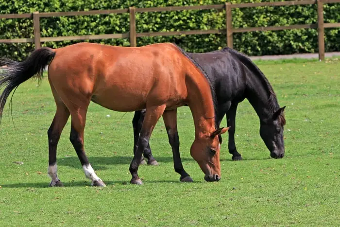
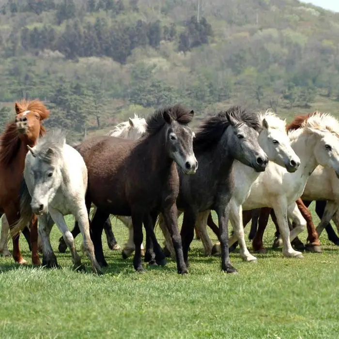
Borna disease is an acute or subacute, non-purulent, often fatal viral encephalomyelitis of solipeds (horses, mule, donkeys) and occasionally of sheep. Borna disease virus can cause a slow infection in horses that affects the central nervous system. This virus is more resistant to environmental influences than other equine encephalomyelitis viruses. The disease and the virus are indistinguishable from Near Eastern Equine Encephalomyelitis (NEEE)
Causes of Borna Disease in Horses
The disease is caused by Borna Disease Virus (BDV) under the family Bornaviridae. The causative virus has not yet been accurately identified. However, the virus is between 85 to 125 nm in size, has a complex structure, and possesses a complement-fixing, soluble antigen (S antigen). The virus is presumed to be a single-stranded RNA type, but its precis structure remains unknown.

Epizootiology of Equine Borna Disease
Borna disease was first described as an epizootic in the Borna region near Leipzig, Germany, in 1894. It remains epizootic in Southern areas of the Federal Republic of Germany, in Hensen, and Switzerland. As a rule, the morbidity of Borna Disease is not high within a given population. There is much seasonal variation in its occurrence. While most cases tend to occur during spring, infections are observed throughout the year. It is not known whether actual reservoirs for the virus exist. It has zoonotic significance also.

Pathogenesis of Fetal Viral Encephalomyelitis
Borna disease is an infectious encephalomyelitis of horses, but natural infection also occurs in sheep. The spontaneous disease is known to occur in rabbits, goats, cattle, and humans.
The disease is spread by inhalation of the virus. The NEEE virus is thought to be transmitted by a tick, probably by Hyaloma spp, and the infection of horses is accidental and outside a normal tick-bird cycle. It is possible that the viruses are the same and that the German disease is created by the periodic transfer of the virus from Near East Europe by migratory birds. The morbidity in Borna disease is not high, but most affected animals die. As a rule, only horses are susceptible.

The viruses appear to have a specific affinity for ganglion cells since the characteristics of intranuclear inclusions, the Joest-Degan bodies, are found in these cells. Current evidence suggests that the virus primarily affects the CNS centripetally along the nerves and then spreads centrifugally to invade the salivary gland, kidneys, mucous membranes, and mammary glands. The most pronounced ganglion changes are found in the olfactory bulb, frontal brain, and hippocampus.
Clinical Findings of Borna Disease in Horses
There are acute, chronic, or persistent forms of the disease. The incubation period is about four weeks. The initial symptoms of Borna disease are non-specific. The signs include:
- Fever
- Anorexia
- Drowsiness.
In the acute form, there is moderate fever, pharyngeal paralysis is seen in terminal stages, and death occurs in 1 to 3 weeks.

In the common chronic form, clinical signs are not evident, but clinical symptoms may occur. Central nervous signs become increasingly prominent as the disease progresses. These includes:
- Incoordination of gait.
- Ataxia.
- Nystagmus.
- Paresis of the limbs.
- Disturbances of equilibrium.
In the field outbreaks, the incubation period is about four weeks and possibly up to 6 months. There is moderate fever, pharyngeal paralysis, lack of feed intake, muscle tremor, and hyperplasia. Lethargy, sleepiness, and flaccid paralysis are soon seen in the terminal stages, and death occurs 1-3 weeks after the first appearance of clinical signs.
Necropsy Findings of Fetal Viral Encephalomyelitis of Horse
At necropsy, there are no gross findings. Histologically, there is typical viral encephalitis, affecting chiefly the brain stem and a lesser degree of myelitis. Diagnostic intranuclear inclusion bodies are present in nerve cells of the hippocampus and olfactory lobes of the cerebral cortex. On death, the brain should be examined for the virus (antigen). The virus may be demonstrated in the cell culture or by rabbit inoculation. In mild cases, the rabbit will contract a lethal meningoencephalitis in 3-4 weeks. Histologically, inclusion bodies in the hippocampus of the caudate nucleus and the olfactory brain will be seen in the ganglion cells.
Diagnosis of Equine Borna Disease
The diagnosis is based on:
- Serological tests and isolation of the virus from the brain of the animals that die or are destroyed.
- Antibody titers in CSF exceed those in serum, and CSF or brain tissues are suitable for diagnosing the disease.
- Tests for serum antibodies are helpful only if positive.
- The CF test, in particular, may be falsely negative in infected animals.
- Indirect Immunofluorescent neurology of the CSF is a better method.
- The CSF tends to contain more significant amounts of antibodies than the serum, especially if the infection has been present for more than 14 days.
Treatment of Borna Disease in Horses
No specific treatment is available. The following treatment and medication can be given for temporary benefits only:
- Brain edema in acute cases can be reduced by IV administration of mannitol (1gm/kg body weight of 10% solution) to induce diuresis.
- A cisternal puncture can achieve the same effect.
- Adrenocortical hormones can be given to reduce inflammation.
Prophylaxis and Prevention of Borna Disease
The preventive measures of Equine Borna Disease are:
- Prophylaxis vaccination should be considered in areas where Borna disease is epizootic.
- The live vaccine of Zwick (Hahn) derived from the brain of an infected rabbit is commonly used.
- Immunity develops following three weeks of subcutaneous injection of 10ml dose.
- Since the BVD vaccine contains a live virus, it should be given only in epizootic areas.
- Foals less than one-year-old and mares in late pregnancy should not be vaccinated.
- Horses between 1 to 2 years old, ponies, and donkeys are given a half dose.
- Horses, sheep, and giants should be housed separately.
- Horses from enzootic areas should be quarantined for two months and tested serologically for specific antibodies in paired tests conducted one month apart.