Melanoma in dogs is a type of cancer originating from the pigment-producing cells called melanocytes. These cells produce melanin, giving skin, hair, and eyes color. When melanocytes undergo uncontrolled growth and proliferation, they form tumors known as melanomas.
Melanoma can affect dogs of any age, breed, or sex, but certain breeds may be more predisposed to this condition. These breeds include Schnauzers, Scottish Terriers, Poodles, and Doberman Pinschers.
Causes of melanoma in dogs
The exact causes of melanoma in dogs are not fully understood, but a few factors may contribute to its development. These contributing factors can vary depending on the type of melanoma (cutaneous or oral) and the individual dog’s genetics and environment. Some potential causes and risk factors for melanoma in dogs include:
- Genetic Predisposition: Certain dog breeds have a higher genetic predisposition to develop melanoma. For example, Schnauzers, Scottish Terriers, Poodles, and Doberman Pinschers are among the breeds with an increased risk of melanoma.
- Exposure to Ultraviolet (UV) Light: Chronic exposure to sunlight and ultraviolet (UV) radiation is associated with the development of cutaneous melanomas in dogs, similar to the link between sun exposure and skin cancer in humans. This is one reason some melanomas occur in body areas with pigmented skin, as these areas may receive more sun exposure.
- Age: Melanomas are more common in older dogs, although they can occur at any age.
- Hormonal Influence: Some studies suggest that hormones may play a role in melanoma development, especially in female dogs with ovarian tumors.
- Immune System Function: A dog’s immune system detects and eliminates abnormal cells, including cancer cells. A weakened or compromised immune system may contribute to the development of melanoma.
- Environmental Factors: Exposure to some environmental toxins or carcinogens may increase the risk of developing cancer, including melanoma.
- Inherited Mutations: In some cases, specific genetic mutations may be passed down through generations and increase the likelihood of developing melanoma.

Types of Canine Melanomas
Canine melanomas are classified based on their location in the body. There are two main types of melanomas in dogs:
- Cutaneous Melanoma:
- This type of melanoma develops on the skin and is the most common form in dogs.
- Cutaneous melanomas can occur in any body part but are often found in areas with pigmented skin, such as the mouth, toes, nail beds, prepuce (sheath in males), and perianal region (around the anus).
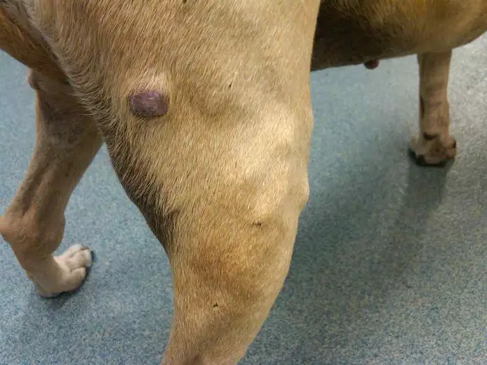
- They may appear as dark or pigmented lumps or masses on the skin.
- Cutaneous melanomas can be categorized into benign (non-cancerous) and malignant (cancerous) forms. Malignant cutaneous melanomas have the potential to spread to other organs of the body, whereas benign melanomas do not metastasize.
- Oral Melanoma:
- Oral melanomas occur in the mouth, particularly on the gums, tongue, and other oral tissues.
- They are often more aggressive and malignant than cutaneous melanomas.
- Oral melanomas are one of the most common malignant tumors in the canine oral cavity and carry a higher risk of spreading (metastasizing) to regional lymph nodes and distant organs.
- They may appear as darkly pigmented, raised masses in the mouth and can cause difficulty eating, drooling, or bleeding from the oral cavity.

Clinical Signs of melanoma in dogs
The clinical signs of melanoma in dogs can vary depending on the tumor’s location (cutaneous or oral) and whether it is benign or malignant. It’s important to note that not all melanomas present the same way, and some dogs may show no apparent symptoms until the tumor has reached an advanced stage. Here are the typical clinical signs associated with melanoma in dogs:
- Cutaneous Melanoma (Skin Melanoma):
- Dark or pigmented lumps or masses appear on the skin, especially in areas with pigmented skin like the mouth, toes, nail beds, prepuce (sheath in males), and perianal region (around the anus).
- These masses may be flat or raised and can have irregular edges.
- Some cutaneous melanomas can resemble other skin growths, making it essential to differentiate them through proper examination and biopsy.
- Malignant cutaneous melanomas may be more aggressive, proliferate, and have a higher likelihood of spreading to other body parts (metastasis).
- Benign cutaneous melanomas typically do not metastasize but should be monitored and treated if necessary.
- Oral Melanoma:
- Oral melanomas are one of the most common malignant tumors in the canine oral cavity.
- They may appear darkly pigmented and have raised masses or ulcers on the gums, tongue, and other oral tissues.
- Difficulty eating or swallowing, drooling, bleeding from the mouth, and bad breath are common signs.
- Oral melanomas tend to be more aggressive and have a higher risk of metastasis, often spreading to regional lymph nodes and distant organs.
- Other Signs:
- Weight loss: Advanced cases of melanoma, especially those with metastasis, can lead to weight loss and general debilitation.
- Lameness: If melanoma occurs on the toes or in the footpad, lameness or limping may be observed.
- Discharge or bleeding: Melanomas can sometimes ulcerate or bleed, leading to discharge from affected areas.
Diagnosis of Canine Melanoma
The diagnosis of canine melanoma involves a combination of thorough physical examination, fine-needle aspiration or biopsy, and sometimes additional imaging or staging procedures. Here is the typical process for diagnosing canine melanoma:
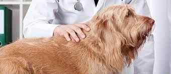
- Physical Examination: The first step is a comprehensive physical examination by a veterinarian. They will carefully inspect your dog’s skin, oral cavity, and other relevant areas to identify any suspicious lumps, growths, or abnormalities.
- Fine-Needle Aspiration (FNA): If a lump or mass is detected, the veterinarian may perform a fine-needle aspiration. During this process, a small needle is inserted into the mass to collect a sample of cells for examination under a microscope. FNA can provide preliminary information about the nature of the mass and help differentiate melanoma from other types of tumors.
- Biopsy: For a definitive diagnosis, a biopsy is often recommended. This involves surgically removing a portion or the entire mass and sending it to a veterinary pathologist for analysis. The pathologist will examine the tissue under a microscope to confirm the presence of melanoma and determine whether it is benign or malignant.
- Staging and Imaging: Further staging and imaging procedures may be required for oral melanoma or suspected metastatic disease cases. This can include X-rays, ultrasound, CT scans, or MRI to assess if the cancer has spread to regional lymph nodes or other organs.
- Histopathology: The biopsy sample will undergo histopathological analysis, where the tissue is examined in detail to determine the tumor type, grade, and stage. This information is crucial for devising an appropriate treatment plan.
- Immunohistochemistry (IHC): In some cases, immunohistochemistry may be used to determine specific markers on the tumor cells, providing additional information that can influence treatment decisions.
Differential Diagnosis of Melanoma in Dogs
The differential diagnosis of melanoma in dogs involves considering other conditions or diseases that can present with similar clinical signs or features. Veterinarians need to rule out these other possibilities to arrive at a definitive diagnosis of melanoma. Some of the main conditions that may be included in the differential diagnosis of canine melanoma include:
- Benign Skin Tumors: Several benign skin tumors can resemble melanomas, such as benign melanocytomas (benign tumors of melanocytes), sebaceous adenomas, fibromas, lipomas, and histiocytomas. Differentiating between benign and malignant tumors is essential for determining the appropriate treatment approach.
- Mast Cell Tumors: Mast cell tumors are common skin tumors in dogs and can appear similar to melanomas in their early stages. These tumors arise from mast cells, immune cells involved in the body’s allergic response.
- Hemangiomas and Hemangiosarcomas: Hemangiomas are benign tumors from blood vessels, while hemangiosarcomas are malignant tumors derived from the same cell type. Both can appear as dark masses on the skin and can sometimes be confused with melanoma.
- Squamous Cell Carcinoma: This is another type of skin cancer that can occur in dogs. Squamous cell carcinomas commonly affect areas of the body exposed to sunlight, such as the nose, ears, and eyelids. Squamous cell carcinomas can be pigmented and may resemble melanoma.
- Oral Squamous Cell Carcinoma: Similar to the cutaneous form, squamous cell carcinoma can also occur in the oral cavity and may have a dark appearance, leading to potential confusion with oral melanoma.
- Lymphoma in Dogs: Lymphoma is a type of malignant tumor affecting lymphocytes, white blood cells in the immune system. In some cases, lymphoma can present as skin masses, leading to a differential diagnosis with melanoma.
- Foreign Body Reaction: Occasionally, the body’s response to a foreign object or material, such as a splinter or suture material, can lead to the formation of a lump or mass that may be mistaken for a tumor.
- Infectious Lesions: Certain infections, such as abscesses or fungal infections, can also form masses that might be confused with tumors.
Treatment of Canine Melanomas
The treatment of canine melanomas depends on several factors, including the type of melanoma (cutaneous or oral), its location, size, whether it is benign or malignant, and the stage of the disease. Treatment options may vary, and sometimes, a combination of treatments may be used. Here are the primary treatment modalities for canine melanomas:
- Surgery: Surgical removal is often the primary treatment for cutaneous and oral melanomas. If the melanoma is small and localized, the goal is complete excision of the tumor along with a margin of healthy tissue. In some cases, reconstructive surgery may be required after tumor removal.
- Radiation Therapy: Radiation treatment may be used in cases where complete surgical removal is impossible or when there is a high risk of local recurrence. It benefits oral melanomas, which tend to be more aggressive and challenging to excise completely.
- Immunotherapy: Immunotherapy is a relatively newer approach in veterinary oncology and involves using medications that stimulate the dog’s immune system to recognize and attack cancer cells. Immunotherapy may be used as a standalone treatment or in combination with other therapies.
- Chemotherapy: Chemotherapy is commonly used in malignant melanomas with a high risk of metastasis (spreading to other body parts). It can help slow down tumor growth and reduce the risk of cancer spreading.
- Targeted Therapy: Targeted therapies are medications that specifically target specific molecules or pathways involved in cancer cell growth and survival. These treatments are becoming more prevalent in veterinary oncology and may be considered in specific cases.
- Cryotherapy or Electrocautery: Cryotherapy (freezing) or electrocautery (burning) may be minimally invasive treatments for small, superficial cutaneous melanomas or early-stage oral melanomas.
- Palliative Care: In cases where the melanoma is too advanced or has spread significantly, palliative care may be the focus. Palliative care aims to manage symptoms, improve the quality of life, and comfort the dog.
Prevention of Melanoma in Dogs
Preventing melanoma in dogs involves a combination of lifestyle choices, early detection, and regular veterinary care. While not all cases of melanoma can be prevented, these measures can help reduce the risk and improve the chances of early detection and successful treatment:
- Limit Sun Exposure: Since UV radiation is associated with the development of cutaneous melanomas, limit your dog’s exposure to direct sunlight, especially during peak hours of the day (typically between 10 am and 4 pm). Provide shaded areas and keep your dog indoors during sunny days, especially if they have light-colored skin or thin fur.
- Sunscreen: If your dog spends time outdoors, particularly in areas with intense sunlight, consider using a pet-safe sunscreen on exposed skin and areas with thin fur. Consult your veterinarian for recommendations on appropriate sunscreens for dogs.
- Regular Veterinary Check-ups: Schedule regular check-ups with your veterinarian, ideally at least once a year and more frequently for older dogs or those with a history of melanoma or other cancers. Regular examinations can help identify any suspicious lumps or skin changes early on.
- Early Detection: Be vigilant about checking your dog’s skin and oral cavity for any unusual growths, lumps, or changes in pigmentation. If you notice any abnormalities, consult your veterinarian for evaluation and possible biopsy.
- Breed-Specific Monitoring: Some dog breeds are more predisposed to developing melanoma. If you have a breed with a higher risk, be especially vigilant in monitoring for any signs of melanoma and discuss preventive measures with your veterinarian.
- Spaying and Neutering: Some evidence suggests that spaying female dogs and neutering male dogs early may reduce the risk of certain cancers, including melanoma. Consult your veterinarian about the appropriate timing for spaying or neutering your dog.
- Environmental Control: Minimize your dog’s exposure to potential carcinogens and toxic substances in the environment. Avoid using harmful chemicals or pesticides in areas where your dog spends time.
- Good Nutrition and Exercise: A balanced diet and regular exercise are essential for overall health and immune function, which can help reduce the risk of cancer and other diseases.
Remember that while these preventive measures can reduce the risk of melanoma, some cases may still occur despite taking precautions. If you have concerns about your dog’s risk or want more personalized advice on melanoma prevention, consult your veterinarian or a veterinary oncologist.
How Long Does a Dog Live With Melanoma?
The prognosis and life expectancy for a dog with melanoma can vary significantly depending on several factors, including the type of melanoma (cutaneous or oral), the stage of the disease at the time of diagnosis, the location and size of the tumor, whether it is benign or malignant, the dog’s overall health, and the treatment options pursued.
Cutaneous Melanoma:
- Benign cutaneous melanomas are usually not life-threatening, and dogs can live an average lifespan with proper treatment.
- Malignant cutaneous melanomas can be more aggressive and may have a higher risk of spreading to other body parts (metastasis). The prognosis for malignant cutaneous melanoma is generally guarded, and survival times can range from several months to a year or more with appropriate treatment.
Oral Melanoma:
- Oral melanomas are generally more aggressive and have a higher risk of metastasis than cutaneous melanomas.
- The prognosis for oral melanoma is generally more guarded, and untreated cases may have a life expectancy of around 1-5 months.
- With aggressive treatment approaches, including surgery, radiation therapy, immunotherapy, and sometimes chemotherapy, survival times may be extended, and some dogs can live for 1-2 years or longer.
Concluding Remarks on Melanoma in Dogs
Melanoma in dogs is a type of cancer that arises from the pigment-producing cells called melanocytes. It can occur on the skin or oral cavity and may be benign or malignant. While not all cases of melanoma can be prevented, there are measures that dog owners can take to reduce the risk and improve early detection.
Early detection is critical to achieving a better prognosis and a successful treatment outcome. Regular veterinary check-ups, vigilant monitoring for skin changes or growths, and prompt evaluation of abnormalities are essential for early identification.
Treatment options for melanoma in dogs include surgery, radiation therapy, immunotherapy, chemotherapy, targeted therapies, and palliative care. The choice of treatment depends on the type and stage of melanoma, as well as the overall health of the dog.
The prognosis for canine melanoma varies widely, ranging from a benign and treatable condition to a more aggressive and life-threatening disease. Benign cutaneous melanomas typically have a good prognosis with appropriate treatment, while malignant oral melanomas have a more guarded prognosis.
Advances in veterinary medicine continue to provide new treatment options and improve the overall care and quality of life for dogs with melanoma. If your dog is diagnosed with melanoma, it’s essential to work closely with your veterinarian or a veterinary oncologist to develop a comprehensive treatment plan tailored to your pet’s needs.
Remember that each case of melanoma is unique, and the information provided here is general. The best action is to consult a qualified veterinarian who can assess your dog’s situation and provide personalized guidance and care. By being proactive in your dog’s health care and seeking appropriate veterinary attention, you can optimize the chances of a positive outcome and provide the best care for your beloved canine companion.
