Mud fever (subcutaneous streptothricosis) is an exudative dermatitis of animals, and very occasionally humans. It is most prevalent in ruminants and only occurs sporadically in horses. With the possible exception of sheep, animals in tropics are more often and more severely affected than those in temperate areas. The condition is sometimes referred to as rain scald, dermatophilosis or greasy heel.
Causes of Mud Fever
The penetration of Dermatophilus congolensis produces the disease into the epidermis. D congolensis belongs to a large, heterogeneous group of bacteria, the actinomycetes, whose primary habitat is the soil. However, D congolensis lives only in the living tissue of the epidermis. It is a gram-positive organism which grows as branching filaments, the hyphal cylinders of which condense in two planes at right angles giving rise to coccoid bodies these coccoid bodies eventually become highly motile zoospores less than1 micrometer in diameter and are the essential means of dissemination of the pathogen. The filamentous mass- the mycelium- readily fragments.
Epidemiology of Dermatophilosis
The only known reservoirs of D congolensis have chronically affected the animal. The organism may be present in a quiescent state in the epidermis until local climate conditions, precisely a period of warm, wet weather, promotes further multiplication of the mycelium and the formation of the zoospores. To establish infection in new areas of skin in an infected animal, or a previously uninfected animal, the motile zoospores, responding to carbon dioxide diffusing through the skin, must penetrate the sebaceous layer and enter the epidermis. They germinate in the dermis, producing a hypha and ultimately a mycelium.
Minor abrasions of the superficial layers of the skin make an animal more susceptible to infection. The disease is commoner in younger animals because their sebaceous layer is thinner and can be more readily weakened by prolonged wetting. Biting arthropods such as ticks and flies, which are more prevalent and active in warm, humid conditions, play a part in the mechanical transfer of zoospores. The extent to which sweating by horses favors infection does not appear to have been investigated.
Clinical Findings and Pathogenesis
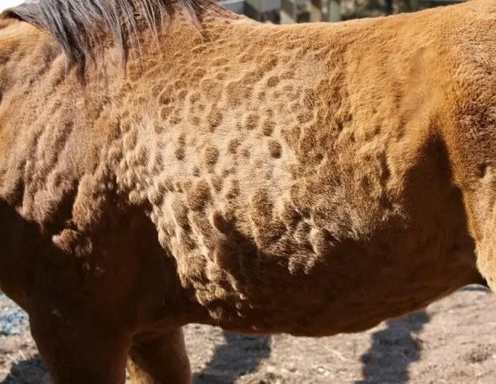
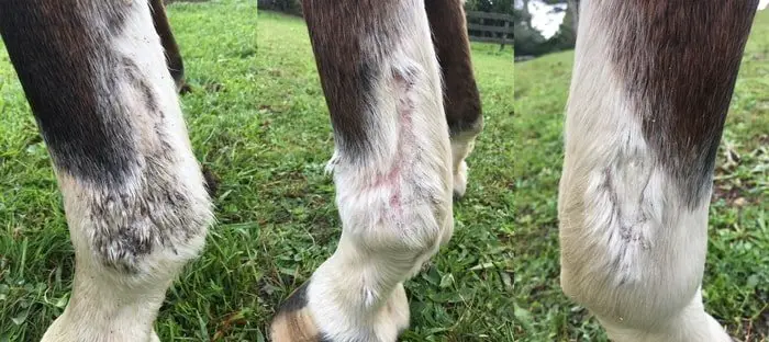
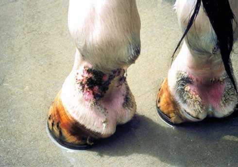
Foals and young horses are the most susceptible. Any part of the integument may be involved although the back, shoulders, and flanks are usually the most affected. Instances in which the patterns and coronets were the main areas affected have also been described as mud fever in horses.
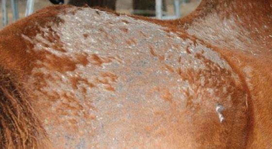
In the early stages after infection, influxes of neutrophils into the affected skin, inflammatory reactions, and the severe resultant exudation mat the hairs of the coat together and produce the characteristic, so-called “Old paintbrush” appearance of the lesions. These lesions, which are scattered over the skin, coalesce with others to create circular, raised crusts or scabs to 2 cm in diameter. They do not itch and most heal spontaneously within a few weeks though a few may persist indefinitely. The factors that govern the curtailment of spread and subsequent healing of the lesions in about 3 wk or less are imperfectly understood.
Diagnosis of Mud Fever
The appearance of the lesions in a clinically affected animal is highly suggestive of streptothricosis, which requires differentiation only from the injuries of the early stages of ringworm or pyoderma.

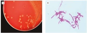
A confirmed diagnosis is early made by observing the unusual morphology of D congolensis in smears of exudate or scabs that have if necessary been moistened, crushed and teased and stained with methylene blue or Giemsa. Preparations stained by fluorescent antibody are useful when the original sporulating structures are scarce, as they are in material from attempted from specimens that contain affected epidermis. Growth of D congolensis in the laboratory is rather slow (taking 2-3 days) but the distinctive colonies, which are often surrounded by a zone of hemolysis, are easily recognized.
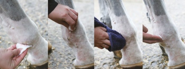
Treatment and Control of Dermatophilosis in Horse
Since the lesions of streptothricosis usually heal spontaneously, therapy is required only for severe cases. Penicillin and streptomycin have a synergistic action on D congolensis in vitro, and a single parenteral injection of a combination of these drugs cures most cases. In severely affected horse in the tropics, an individual does of long-acting oxytetracycline is highly effective.
Since cases of cutaneous streptothricosis in the horse are mostly sporadic and much dependent on weather conditions, there has been no requirement for protection by immunological means. In any case, variability among the antigens of the flagella of the zoospores of D congolensis makes the prospects for a universally applicable vaccine unlikely.

