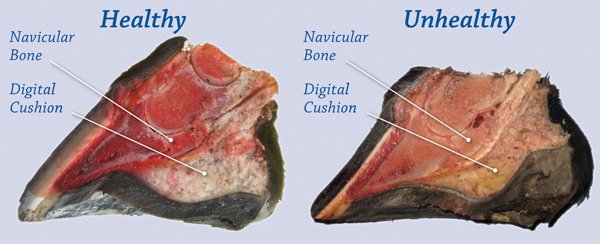
The navicular bone is a small fat bone that lies across the back of the coffin bone of horse hoof. Navicular bone horse is attached to the pedal bone by a strong ligament and the pastern bone by a suspensory ligament. There are several soft tissue structures are associated with navicular bone. Fractures can occur as a result of direct trauma to the foot from the hard or stony ground during exercise, from kicking a solid object, or from internal trauma due to abnormal stresses. Fractures can also occur secondary to advanced navicular disease changes.
What is the cause of navicular bone in horses?
The most common cause of navicular bone in the horse is the heel pain. There are many theories point to abnormal conformation and foot imbalance as the main predisposing factors in the development of the condition. Abnormal foot confirmations implicated are:
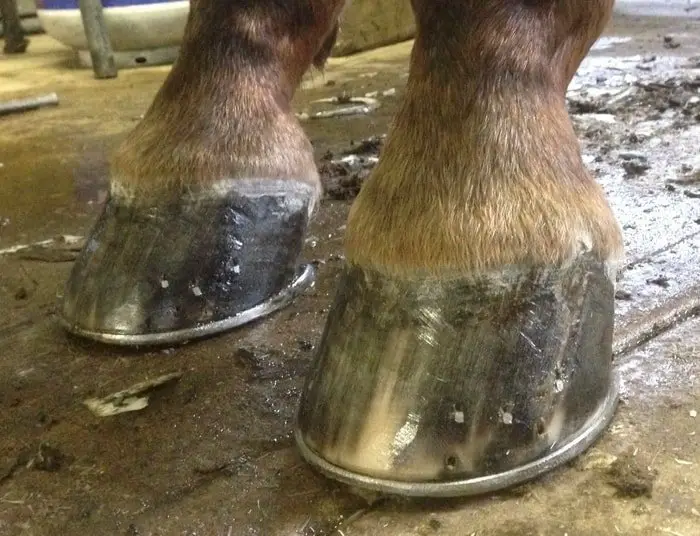
- Backward broken hoof-pastern axis: this is probably the most common, and is almost always the result of long toe/low heel imbalance
- Feet smaller than normal in reaction to body size
- Mediolateral foot imbalances
- Sheared heels
What are the Clinical Signs?
There is usually a history of a sudden onset of severe lameness. In some cases, the horse will only bear weight on the toe of the foot. Usually, there will be a marked pain reaction to the flexion of the DIP joint. A pain response can often be elicited by pressure with hoof testers across the posterior third of the hoof wall. After 2-3 wk the degree of lameness usually lessens but can be exacerbated by flexion of the DIP joint or exercise.

How is Navicular Bone Horse diagnosed?
The diagnosis is based on history and clinical signs. A palmar digital nerve block will often improve the lameness, however, a negative response to the nerve block does not rule out a fracture of the navicular bone.
The diagnosis can be confirmed by radiography. Great care should be taken in the preparation of the foot before the radiographic examination, and the sulci of the frog should be packed so that radiolucent lines from the air are not mistaken for fractures. At least two radiographic projections demonstrating the fracture line are necessary before a definitive diagnosis is made.

Although rare, congenital bipartite and tripartite navicular bones have been reported. These may not cause lameness and can be distinguished from fractures radiographically by their smooth rounded edges, and the overall symmetry of the bone.
How Do You Treat Navicular Bone Horse?
Conservative methods involved prolonged rest (6-8 months), combined with hoof trimming and balancing of the foot and shoeing with a full bar shoe which does not contact the frog, fitted with quarter clips and a rolled toe. Alternatively, trimming and balancing the foot and shoeing with a graduated raised heel shoe, fitted long in the heels and with quarter clips, can be tried. The horse is given 2 months box rest followed by a gradually increasing, controlled exercise program.
Whatever method of conservative treatment is used, healing will be slow and by the fibrous union because of the difficulty in immobilizing the fracture. The fracture line will always be evident radiographically and complete soundness is uncommon.
Surgical treatment by lag screw fixation can be successful in the treatment of navicular bone fractures and results in a bony union without excessive callus formation. Repair should be performed as soon as possible after the fracture has occurred to reduce secondary changes. The surgical technique is complicated and sophisticated equipment is required.
Concluding Remarks on Navicular Bone
Horses of all foot shape can develop navicular bone problems. Balanced ration, correct shoeing and maintaining good stable management can reduce the incidence of the condition. Though the prognosis is poor in the case of navicular bone problems, careful management can reduce the sufferings of your horse. As a horse owner, you must take care of the causes of navicular syndrome.