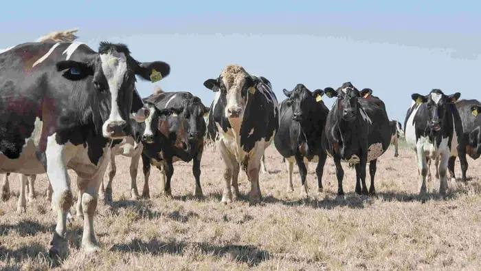
Theileriosis in cattle is a tick-borne disease caused by haemoprotozoa Theileria spp. The disease is manifested by high fever, enlarged lymph nodes, severe anemia, and death in some infected animals. The disease is highly endemic in South Asian countries. The Theileria infected cattle are ubiquitous in distribution and are the main reservoir of infection. Theileriosis has been recorded in animals of all ages. Calves are equally susceptible but are very mild in adult indigenous cattle. Though the indigenous cattle suffer only in appearance, they cause extensive damage to exotic cattle and their crossbreeds by being asymptomatic carriers of this infection.
Causes of Theileriosis in Cattle
Theileriosis is a disease of cattle caused by a blood protozoan parasite Theileria of which the following species are common.
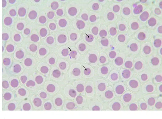
- Theirelia Parva is highly pathogenic and causes East Coast Fever (ECF). The organism is a rod and comma-shaped but pleomorphic during a recovery phase. Approximately 80% of erythrocytes are seen parasitized with 1 to 12 organisms. Immunity persists after natural infection. The cross-immunity with T lawrenci only and not with others.
- Theileria annulata is moderately to highly pathogenic, lethal for cattle but mild for buffaloes. The organism causes Mediterranian Coast Fever or Tropical Theileriosis. The erythrocytic forms are predominantly oval or annular.
- Theileria mutans piroplasma is pleomorphic rods, 2.0 to 5.0micron, long numerous of the first infection, rare in blood for years. Schizonts are absent within the RBC.
- Theileria lawrenci causes a highly fatal disease in cattle called Corridor or Rhodesian theileriosis.
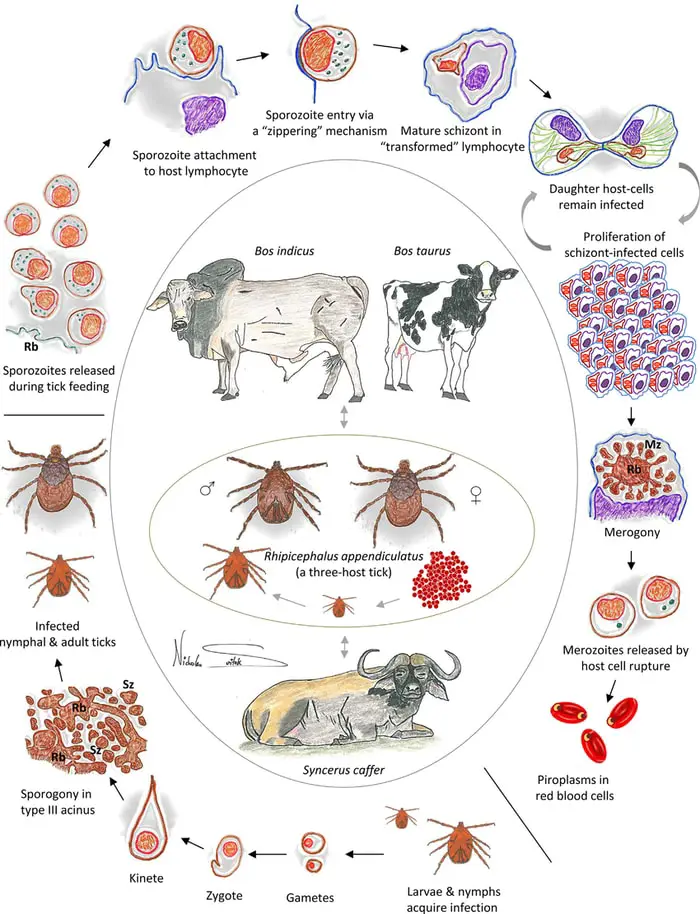
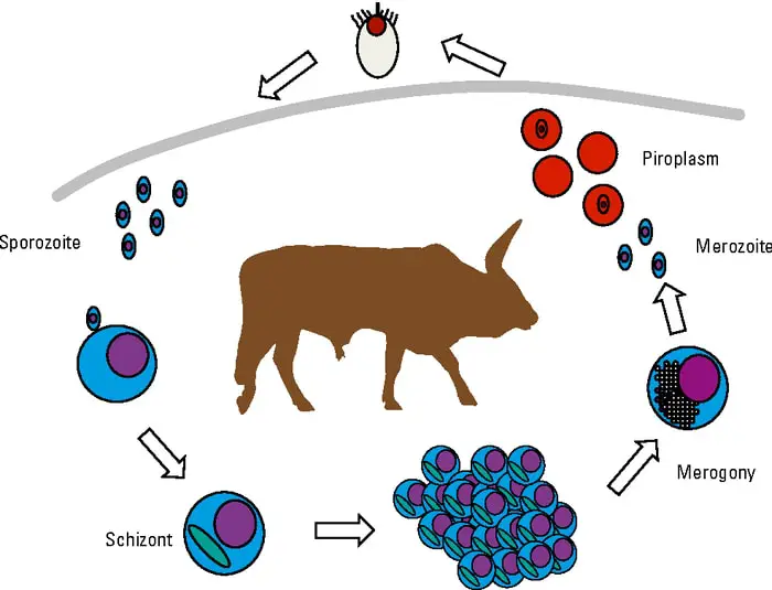
Life Cycle of Theireria
- The Theileria parasite passes through three distinct stages in its life cycle.
- The sporont stage occurs in the ticks, followed by schizogony in the mammalian host when the infective particle with the vector’s saliva is injected into the cattle body by the tick during feeding.
- Lymphopoeisis and enlargement of lymph nodes take place.
- Multinucleated bodies known as KOCH’S BLUE BODIES are found in the cytoplasm of lymphocytes at this stage.
- Later in the infection, a different form of multinucleated microschizonts is formed in the infected lymphocytes, reticulated cells, or macrophages.
- Micromerozoites are formed from these, which coincide with the piroplasmic stage.
- These parasites are ingested by the larval and the nymphal ticks and blood while feeding on the infected animals.
General factors Regarding Tick Transmission
- The biologically transmitted infection causes more severe disease than a mechanical one.
- The process of tick molting is essential for the parasite to reach salivary glands and making the tick infective. Adult tick fed on infected animals when attached to another susceptible animal does not transmit the infection.
- When a tick has molted, feeding is essential for the final development of the tick and parasite.
Pathogenesis of Theileriosis in Cattle
There are two distinct types of pathogenicity involved in theileriosis:
- Induced by macroschizonts.
- Induced by erythrocytic forms.
Pathogenicity of the disease in T Parva infection is mainly related to macroschizonts, lymphoid tissue replication. In T annulata, it is induced both by macroschizonts and well by piroplasmic forms. In T mutans, it is only due to piroplasmic forms.
Clinical Signs of Theileriosis in Cattle
The disease’s incubation period varies from 8 to 12 days, and the course of the disease lasts for 10-19 days.
- The first sign is an enlargement of lymph nodes draining the site of tick feeding.
- Fever commences one to four days later.
- There is usually an abrupt rise of temperature to 103 to 104 F.
- There may be slight remission in fever but again rises, reaching 105 to 106 F within two days of its commencement and remaining high until death.
- There is loss of milk production and malaise, and swelling of all the superficial lymph glands.
- The appetite is reduced, and there is a progressive loss of condition.
- Respiration is accelerated and accompanied by a short spell of coughing.
- Initially, there may be constipation, but after few days of fever, diarrhea is usually present.
- The feces contain blood and have a coating of bloody mucous.
- Leukopenia is seen in T parva infection.
- There is watery discharge from the eyes during most of the courses of infection, and in some animals, photophobia and peripheral opacity of the cornea develops.
- Clinical anemia is a consistent syndrome with T annulata infection, bilirubinemia, and bilirubinuria, and at times, clinical jaundice is also present.
- A subacute form of the disease occurs in calves born of immune dams in an endemic area. The clinical signs are similar to the acute form but not as pronounced, and recovery is more common.
Post Mortem Lesions of Bovine Theileriosis
Gross lesions were found in fetal cases on the course of the disease and the strain involved. These are mainly related to lymphopoiesis and vascular systems.
- In per-acute cases, the spleen and lymph nodes are small and depleted of lymphoid tissues.
- However, in other cases, there is lymphoid hyperplasia with enlargement of the spleen.
- Petechial hemorrhages of serous and mucous membranes are usually present.
- The liver is enlarged, and the color changes to purple or blue, which is due to interstitial production of lymphoid tissue and in part due to degeneration of parenchyma.
- Lungs are congested and edematous, and interlobular septate may be distended with a jelly-like fluid and forth in small bronchioles.
- Abomasum has small superficial hemorrhagic punched ulcers with a rough or triangular outline.
- Petechial hemorrhages are present over most of the serous mucous surfaces of the entire small and large intestine.
- Splashy hemorrhages may also be seen in the endo and epicardium, especially of the left ventricle.
Diagnosis of Theileriosis in Cattle
The diagnostic process of theileriosis is:
- Clinical signs.
- Blood smear examination.
- Lymph node biopsy.
- Histopathological examination.
- Serological tests like Indirect Fluorescence Antibody (IFA), Complement Fixation Test (CFT), Capillary Tube Agglutination Test (CAT), HI, IHA, Conglunating Compliment Antibody Test (CCAT), and peroxidase test can also be employed for the serological diagnosis of the disease.
- Demonstration of Kock’s Blue Bodies in blood smear and lymph node smear is confirmatory.
- The presence of punched ulcers in the abomasum is a pathognomic lesion.
Treatment of Bovine Theileriosis
The treatment of the disease are as follows:
- Tetracycline 5-10mg/kg body weight IV 7-10 days course.
- Chlortetracycline 15mg/kg body weight parenterally for 8-10 days.
- Oxytetracycline 10mg/kg body weight IM a course of 4 injections with a Terramycin 20mg/kg as a single dose treatment.
- Berenil 10mg/kg body weight IM on an alternate day 3-4 injections.
- Nivaquine 1-15 mg/kg body weight IM for 3-4 injections.
- Halofuginone, an anti-protozoal substance at a dose of 1.2 mg/kg body weight orally, is also highly effective.
- Supportive therapy with antihistaminics, haematinic mixture, and blood transfusion is helpful.
Immunization Against Theileriosis in Cattle
Many immunization techniques against bovine tropical theileriosis have been attempted, but none are entirely successful. Some of the immunization methods are:
- Vaccination with tissues like infected blood or lymph nodes from animals suffering from the disease.
- Vaccination with tissue culture attenuated infected lymphoblast cell that is macroschizonts.
- Vaccination by use of infection and treatment method
- Inoculation of tick’s GUTS (Ground Up Ticks Supernatants)
- Vaccination with schizont adopted in small animals.
Prevention and Control of Bovine Theileriosis
The preventive and control measures of theileriosis are as follows:
- Control of ticks combined with control of cattle movement.
- The strategic use of vaccination.
- Specific chemotherapy where this can be effectively applied.
- Since ticks act as a vector, it is necessary to keep animals in tick-free sheds and surroundings.
- Systemic dipping and rotation of grazing land will be of great help.
- Exotic animals should not have access to pasture or water reservoir uses for indigenous stocks.
- Recording of temperature at regular intervals.
- Examination of blood and lymph nodes of suspected animals.
- Proper management and cleanliness of herds.
- A regular treatment of cattle with suitable acaricide during the period when preimaginal stages of ticks occur.
- Where two host ticks are involved, effective treatment with acaricide every 1-12 days will prevent the development of nymphs and kill all preimaginal stages.
- Where host ticks are involved, 5-7 days of treatment will prevent engorgement.
- A high volatile acaricide should be used for building to attack ticks hidden in cracks or crevices.
- All bushes should be burnt and the area cleared of boulders.
Final Advice on Bovine Theileriosis
Theileriosis is one of the essential protozoan diseases of domestic animals. The organism is present in the erythrocytes and causes high fever and anemia. The organism is difficult to treat and control. The preventive measures can reduce the loss of animals and save your farms. In my article, I have discussed most of the critical points briefly. I think this will help you to prevent and control theileriosis in cattle effectively.