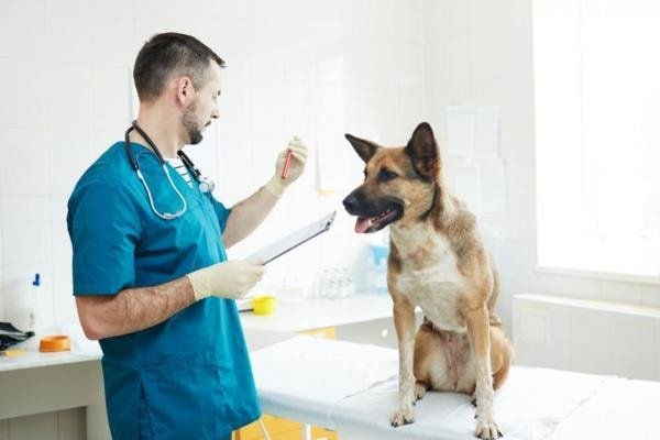
The liver is an essential and vital organ for your dogs. The organ removes the toxic substances from the body from his system. It helps with the process of blood clotting and digestion. Liver disease in dogs is a significant topic for a dog owner. The disease can recover from damage and be treated and managed. You should be given the proper treatment as early as possible, and the recovery depends on the exact cause of the liver damage.
Most Common Liver Disease in Dogs
Your dog may be affected by many diseases and the liver is one of the most important organs that you must be cautious. There are different types of Liver disease in Dogs. These include- acute hepatitis and acute hepatocellular degeneration, chronic inflammatory liver disease, cirrhosis, cholangitis or Cholangiohepatitis, drug-induced liver disease, other hepatobiliary obstruction, gallstones, and liver tumors. The causal agent of liver diseases is many.

1. Acute Hepatitis and Acute Hepatocellular Degeneration
Acute hepatitis and acute hepatocellular degeneration is focal or diffuse damage to the hepatocyte. The causes of hepatocellular disruption of your dog are many and are often undermined. Some of the recognized hepatotoxins include chemical solvents like carbon tetrachloride (CCl4) and mycotoxins like aflatoxins. Your dogs show variable signs include inappetence, lethargy, and vomiting. Icterus may develop if the hepatocellular insult is extensive of your pets.
The predominant biochemical abnormality is an increase in serum ALT, the magnitude of which depends on the severity and extent of the hepatocellular damage. The serum AP is usually normal or only slightly increased early in the disease process. A more severe insult may also increase serum AST. An increase in serum bilirubin concentration may occur if a sufficient number of hepatocytes have been damaged or destroyed or there has been sufficient damage to cause cholestasis.

The patient will recover if enough functional liver remains to support regeneration. A liver biopsy is often neither helpful nor essential in the diagnostic evaluation and maybe contraindicated by liver associated coagulopathies. The histologic examination can be useful for assessing the severity and extent of the disease process of your dog. A liver biopsy is justified if recurrent abnormal ALT AST values have been documented.
The disease can be a differential diagnosis of acute pancreatitis, infectious canine hepatitis, acute gastroenteritis, severe tryptic enteritis, and some toxicosis. Successful recovery depends on aggressive therapy, and few hepatotoxins have specific antidotes. The prognosis of your dog is usually good if the inciting cause is removed, permanent loss of functional mass is less than 50%, and the liver has retained the capacity to regenerate.
2. Chronic Inflammatory Liver Disease in Dogs
Your dog suffers from a variety of chronic inflammatory liver diseases. All have similar clinical signs. In your dog, chronic hepatitis has been associated with leptospirosis, the administration of primidone, phenytoin, and phenobarbital, abnormal copper metabolism in Bedlington Terriers and West Highland White Terriers, and experimentally induced infectious canine hepatitis. A syndrome of chronic hepatitis has also been recognized in your Doberman Pinscher and American Cocker Spaniel.
The disease has overlapping histologic appearances and is mostly of unknown causes. The signs are nonspecific. Inappetence and lethargy are common, but vomiting, diarrhea, and pica may also be seen occasionally in your pets. Icterus may develop terminally. Signs may be chronic, and the disease far advanced before a diagnosis is made.

The disease is characterized histologically by periportal inflammation and fibrosis. More ominous histologic features suggestive of progression are implied when confluent areas of necrosis have identified that zones of parenchymal collapse that bridge between portal triads and central veins or span lobules between portal triads.
The disease is differentiated from infections, drug-induced, familial, or lobular dissecting hepatitis, idiopathic chronic hepatitis. The specific treatment is unavailable in most cases and usually depends on the inciting cause. You should give supportive therapy, including vitamin E, ursodeoxycholic acid, and possibly S-adenosylmethionine(S-AME). Anti-inflammatory therapy using prednisolone, and colchicine may also be useful for your dog.
3. Liver Cirrhosis in Dogs
Cirrhosis is a diffuse hepatic disease process characterized by fibrosis and an alternation of typical hepatic architecture by structurally abnormal regenerative nodules. Your dog needs almost 20% of normal liver function to survive. Cirrhosis is the final, irreversible end-stage of many chronic liver diseases.
Cirrhosis can cause as a result of damage to the liver by many diseases, toxins, or drugs. Different types of diseases that may lead to cirrhosis include cancer as well as bacterial, viral, and fungal infections that cause hepatitis. Some toxins and some long-term use of medications, such as corticosteroids and common pain-relieving medications, can also cause cirrhosis of your dog.

The symptoms may include inappetence, ascites, and mental depression that can progress to your dog to encephalopathy. Sometimes you may be seen weight loss, diarrhea, vomiting, swollen abdomen, bleeding problems, depression, behavior changes of your dog. In this condition, your expert performs a thorough physical examination of your lovely pets.
Biochemical findings are often subtle. Serum ALT and AP activities may be normal or only slightly increased because of the decreased hepatic cell mass. Other biochemical abnormalities reflect the liver’s reduced functional capacity and altered hepatic blood flow secondary to changes in architecture. Decreased serum urea nitrogen and decreased serum albumin concentration may be noted. Increased serum bilirubin concentration is a lagging indicator of liver insufficiency. Persistent bilirubinuria will be detected before jaundice develops.
Determination of total serum bile acids is the most reliable test for detecting hepatic insufficiency in the anicteric patient. Histologic confirmation of cirrhosis is essential since, on gross visual inspection, cirrhosis may resemble metastatic disease or nodular regeneration. You should need supportive measures to control the complications of chronic liver failure, such as hepatic encephalopathy and infection.
4. Cholangitis or Cholangiohepatitis
Cholangitis (inflammation of the bile ducts) and Cholangiohepatitis (inflammation of the adjacent hepatic parenchyma) is a relatively common liver disease of the cat. Still, it is diagnosed with a lower frequency in the dog. The cause is unknown but is believed to be due to ascending bacterial infection of the biliary tract, especially gram-negative and anaerobic organisms—cholestasis results in direct and persistent bile duct damage by lithocholic acid and other secondary bile salts.
Jaundice often develops your dog by that time; the liver disorder is recognized. An acutely painful abdomen may be detected on palpation in your dog, especially if the gallbladder is inflamed (cholecystitis). Early laboratory findings of Cholangiohepatitis include increased ALT, AST, and AP activities and, later, a conjugated hyperbilirubinemia.
Primary treatment includes antibiotics based on the results of culture and sensitivity testing. In chronic disease, glucocorticoids will also be required for your dog. Vitamin E and S-AME therapy may also be used, and nutritional support is an integral part of treating this disease.
5. Drug-Induced Liver Disease
Drugs can cause liver injury ranging from a transient asymptomatic increase in serum transaminase activity to clinically overt acute or chronic liver disease. The causes are drugs that can induce liver injury by either direct toxic action or indirectly via a hypersensitivity reaction. Your dogs may suffer sedation, ataxia, weight loss, anorexia, behavioral changes, coagulopathy, jaundice, and ascites.
A hypersensitivity response is complicated to prove since challenge exposes the patient to unnecessary risk. Sulfa -containing drugs, anticonvulsants, anthelmintics, halothane, and others- has been implicated in your pets’ experts.
It is prudent to discontinue the inciting drug. If the drug is essential, then the dose should be modified as far as possible. Fasting and postprandial bile acid concentrations should be determined, as significant hepatic damage is less likely if the concentrations are average.
6. Extra Hepatobiliary Obstruction
Extrahepatic biliary obstruction refers to the impairment of bile flow in the biliary system between the liver and the duodenum. Acute, total extrahepatic impairment of bile flow is rare. Chronic pancreatitis and tumors of the pancreas and the bile duct epithelia are the most common causes, although gallstones in the common bile duct have been reported in your dog. Inspissated bile (most often associated with an underlying liver disorder) and liver flukes are the additional cause of the disease.

Your dogs may suffer inappetence, icterus, and repeated vomiting when your dog’s pancreas is involved. Others include anorexia, lethargy, and jaundice. Physical findings are usually unremarkable for icterus. If your dog with suspected pancreatitis remains icteric beyond 10-14 days of symptomatic medical management, and extrahepatic component should be suspected and managed accordingly.
Extrahepatic cholestasis causes a marked increase in the hepatic synthesis of AP, wrestling in a concomitant increase in serum activity. Retained bile acids also cause hepatocellular damage with an associated mild to moderate increase in serum ALT activity. Clothing studies reveal increased PT and PTT, which should revert to normal with vitamin K therapy. The test for bilirubinuria will be strongly positive.
Treatment should be performed by surgical correction. Your vet will manage the condition. If no obstruction is found at laparotomy, the liver should be biopsied, and the patient managed accordingly. Ursodeoxycholic acid and vitamin E are also helpful.
7. Liver Disease in Dogs: Gallstones
Gallstones are occasionally found in dogs and cats. Most are serendipitous findings on abdominal radiographs or at necropsy. On rare occasions, gallstones can obstruct the common bile duct, but only finding them on radiographs in patients with evidence of liver disease is not just for removal.
Ionized calcium may play a part in the formation of gallstones as it is the main component of pigment gallstones. Choleliths are rare in dogs owing to the absorption of ionized calcium from the gallbladder, which reduces the concentration of free ionized calcium in bile. Gallstones often cause no signs of your dog and are incidental findings at necropsy or during imaging. If signs are apparent, they may include vomiting, icterus, anorexia, fever, and abdominal discomfort.

Gallstones are diagnosed by abdominal ultrasonography by an expert vet. They appear as hyperechoic foci or can be detected by acoustic shadowing originating from the gallbladder. Exploratory laparotomy should be performed in your vet, and the patency of the common bile duct ascertained if signs persistent. The usual treatment is surgical excision with follow-up therapy with ursodeoxycholic acid.
8. Liver Disease in Dogs: Tumors or Cancers
Primary and metastatic tumors occur in the liver. Metastatic tumors are reportedly twice as frequent as primary tumors. Primary tumors include bile duct adenoma (adenocarcinoma), hepatocellular adenoma, lymphoma, and carcinoma (hepatoma). Hepatocellular carcinomas have been associated with hypoglycemia in the dog. Lymphosarcoma is the most common liver tumor of your dog.

The exact cause of the disease is not usually determined. Potential causes include nitrosamines, liver flukes, aflatoxins, and radioactive compounds. Metastatic tumors of your dog arise from the pancreas, mammary glands, bone, lungs, thyroid, GI tract, and spleen. The signs include inappetence progressing to anorexia, weight loss, vomiting, abdominal distension, and terminal jaundice.
Those tumors which do not cause extrahepatic biliary obstruction may increase serum AST, ALT, AP activities, and total serum bilirubin concentration. Abdominal radiographs are often read as usual, but they may reveal hepatomegaly. Ultrasound-guided biopsy or laparotomy and biopsy are essential for the diagnosis of neoplastic disease of your dog. The gross appearance of neoplastic lesions may appear similar to nodules associated with cirrhosis.
Primary tumors confined to a single lobe may be resected surgically. The abdominal cavity should be evaluated for metastatic spread, and biopsy specimens of hepatic lymph nodes should be obtained. Chemotherapy may be appropriate in your selected patients.
Concluding Remarks
Liver disease in dogs is common, and you will face the problems frequently. The proper management and care you can prevent the diseases in your dogs. Infectious diseases can be treated with antibiotics and liver tonics. Healthy liver resembles a healthy dog. The liver is the most critical organ of the body that is essential for many functions of any animal. I this article, I have discussed the most common liver diseases for your better understanding. I think this article will help you to keep your lovely pet healthy and fit.