Coccidiosis in chickens is one of the most critical protozoan diseases of poultry, caused by Eimeria spp and is characterized by enteritis and bloody diarrhea. Coccidiosis is the most common disease that causes the most significant economic damage to poultry farms. The disease develops irrespective of age, sex, and breed of chickens, but the outbreaks are common in birds of 3-6 weeks of age.
Most Important Information on Coccidiosis in Chickens
Coccidiosis is a common disease in the poultry industry. The annual worldwide cost due to Coccidiosis is estimated at 800 million dollars, including the costs of prophylactic in-feed medication, treatment, and losses due to morbidity and mortality and low feed conversions of birds that survive outbreaks.
What Causes Coccidiosis in Poultry?
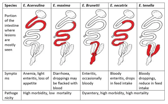
The etiology of the disease is by the protozoa of the Phylum Apicomplexa, family Eimeriidae. Nine species of Eimeria- Eimeria tenella, Eimeria necatrix, Eimeria Brunetti, E. maxima, E. mites, E. acervulina, E. mivati, E. praecox, and E. hagani have been described from chickens, but some are questionable.
Epidemiology of the Disease
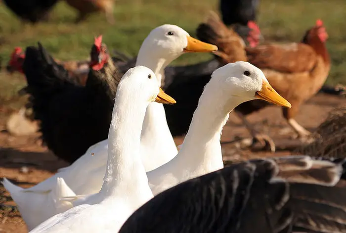
Coccidiosis affects most species of poultry, waterfowl, game birds, and pigeons. However, this disease is almost universally found wherever chickens are raised but is most important where birds are housed or confined in small areas. All domestic birds are susceptible, but the Eimeria species are in the general host, tissue, and cell-specific. Most birds in a flock become infected, but only minority cases develop clinical disease.
Transmission of Coccidiosis in Poultry
Droppings containing oocysts of coccidia are the primary means of transmission of Coccidiosis amongst susceptible chicks. However, mechanical transmission by personnel, equipment, or by wind spreading poultry house dust and litter over short distances is also possible. Both diseased and recovered birds shed oocysts in their droppings, which under conditions of warmth and moisture, develop to the infective stage of sporocyst.
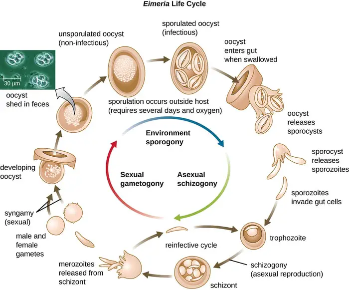
Life Cycle of Coccidiosis in Poultry?
The coccidial life cycles are self-limiting; if new oocysts are not swallowed, the original organisms will have developed through all the endogenous stages. The host will be freed of infection.
- The life cycle of E. tenella is typical of all Eimeria, although some species vary in the number of asexual generations and the time required for each developmental stage.
- The life cycle is initiated when a host swallows a sporulated oocysts.
- After the oocysts, the wall is crushed in the gizzard, and the sporozoites are released in the gut also by the action of pancreatic juice and trypsin.
- The sporozoites enter cells in the mucosa of the intestine and begin the cell cycle leading to reproduction.
- Nearly all species develop in the intestinal epithelium, where each species has a market preference for development in specific sites.
- The free sporozoites invaded the surface epithelium and are engulfed by macrophages in the lamina propia, and are transported into the glands of Lieberkutin.
- They leave the macrophages and enter the epithelial cells lining the glands, and develop into asexual schizonts.
- Intracellular sporozoites undergo nuclear and cytoplasmic changes, which result in asexual multiplication by schizogony. After the schizonts mature, the merozoites are released by rupture of the epithelial cells.
- These merozoites may initiate 2nd subsequent generations of schizonts in the new epithelial cells.
- At least two generations of asexual development, called schizogony or merogony, lead to a sexual phase, where small, motile microgametes (male) seek out and unit with macrogametes (female).
- The resulting zygote matures into an oocyst released from the intestinal mucosa and is shed in the feces.
- A single oocyst may give rise to as many as 100000 progenies.
- The entire process takes 4 to 7 days, depending on species, although oocysts maybe shed several days after patency is reached. In E. tenella and E. necatrix, the maximum tissue damage may occur when second-generation schizonts rupture to release merozoites.
- Other species may have small schizonts, which cause little dame, but the gametocytes may elicit a strong reaction with cellular infiltration and thickened inflamed tissues.
Pathogenesis of Coccidiosis in Chickens
- Harmful effects on the host are primarily done due to the rupture of the epithelial cell lining.
- There is denudation of mucosa, tissue damage, rupture of capillaries, and blood loss.
What are the Signs of Coccidiosis in Chickens?
Several factors influence the severity of the infection, and some of these are the age of the bird and the nutritional status of the host. Young birds are generally more susceptible than older ones. The signs include-
- Bloody diarrhea.
- Dehydration.
- Anorexia and decreased body weight.
- Reduced egg production.
- Infected birds are listless, have ruffled feathers, and reluctant to move.
- Anemia is also common.
- Mortality may be as high as 50% in caecal Coccidiosis.
- In intestinal Coccidiosis, the mortality rate may reach up to 25-30%.
Pathological Lesions of Coccidiosis in Poultry
- In caecal Coccidiosis, the caecal pouch is greatly enlarged and distended and contains clotted blood with mucosa pieces in the lumen.
- The caecal wall is edematous and considerably thickened, while the caecal core may be detached from the mucous membrane.
- In intestinal Coccidiosis, reddish-black areas are seen around the intestine while the intestinal wall has white spots, thickened, distended, and looks flabby.
- Its mucosa may be denuded, and there may be purulent or diphtheroid inflammation.
Diagnosis of Coccidiosis
A tentative diagnosis of Coccidiosis can be made on young birds’ history and recent flock exposure to a large source of sporulated oocysts, clinical findings, and post-mortem changes. Your veterinarians perform a confirmed diagnosis. Post-mortem and laboratory diagnosis are required to confirm the diagnosis. Direct inspection of oocyst in the feces is the other method of diagnosis.
The disease may be confused by- bacterial enteritis, necrotic enteritis, Ranikhet disease, and Rotavirus infection.
How Do You Treat Coccidiosis in Chickens?
A significant advance in the therapy of Coccidiosis in poultry was the introduction of Sulphonamide. Since then, several other anticoccidials have been used successfully. The disease is effectively treated with amprolium in both chickens and turkeys. You must have to increase vitamin A and K in feed or water may reduce mortality and hasten recovery, respectively.
Prevention of Coccidiosis in Chickens
Use of dry litter, turning of litter, and regular changing of litter must e adopted to prevent coccidial infection. Special care is needed in the rainy season when the air’s moisture content remains high along with a suitable temperature that favors the sporulation of oocyst. Anticoccidial drugs should be used in the feed, but their use is not a substitute for the hygienic measures, and new anticoccidial vaccines may be used in layer and breeder flocks.