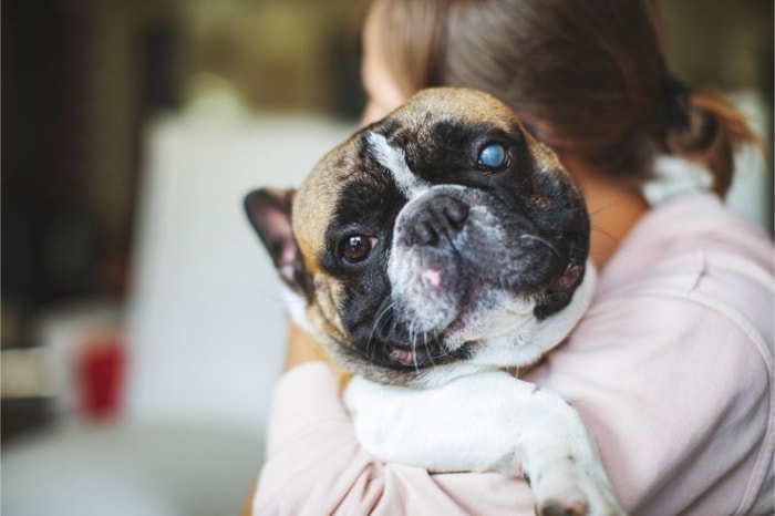
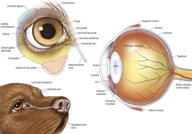
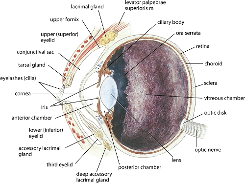
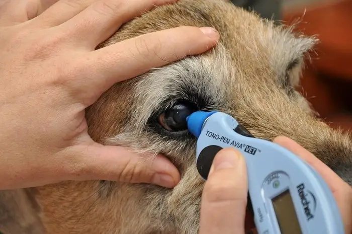
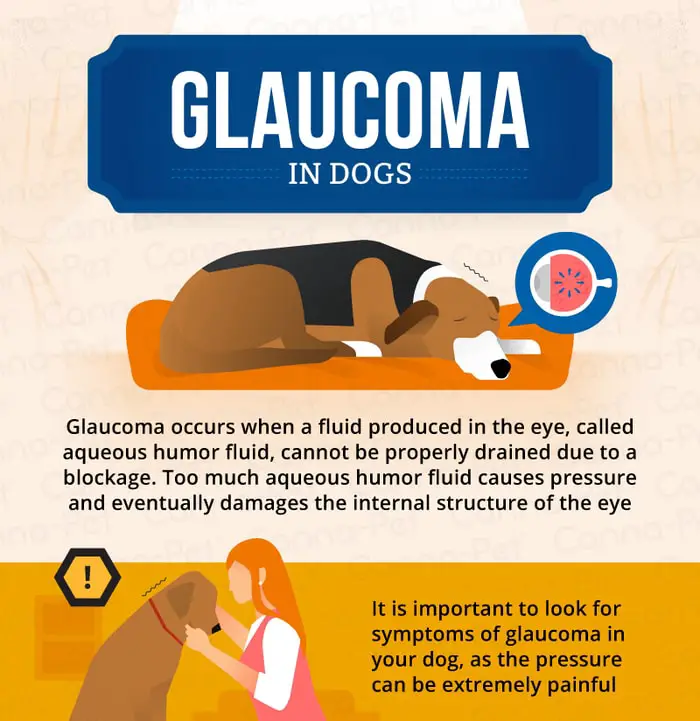
Glaucoma in dogs encompasses a group of diseases characterized by a pathological increase in intraocular pressure (IOP) due to the damage to the optic nerve and retina and vision loss. The intraocular pressure is measured using an instrument called a tonometer. If the condition remains untreated, permanent blindness is an inevitable outcome.
Can a Dog Live With Glaucoma?
Glaucoma is a common disease of the eye in dogs. The disease is not life-threatening. If your dog is diagnosed with glaucoma, it can live its everyday life. The condition should be treated as soon as possible to turn into permanent blindness. The ages dogs and few dog breeds are more susceptible to this condition. As a dog owner, you should have a piece of minimum knowledge on this disease. In my article, I shall discuss the most worthwhile causes and types of glaucoma in dogs.
What Causes Sudden Glaucoma in Dogs?
The inadequate drainage of aqueous fluid causes it, but glaucoma is not caused by fluid overproduction. Glaucoma is classified as either primary (with no other ocular damage preceding glaucoma development) or secondary (where glaucoma develops because of other ocular diseases).
Types of Glaucoma in Dogs
The condition of glaucoma may different types basin the cause, location, and symptoms. The most common types are discussed below:
Primary Glaucoma
Primary glaucoma results in increased intraocular pressure in a healthy eye. Both eyes are affected, although the disease’s clinical signs usually develop in each eye at different times. Primary glaucoma occurs due to inherited anatomical abnormalities in the drainage angle. Primary glaucoma is further divided into the morphological appearance of the iridocorneal angle.
Narrow-angle Primary Glaucoma
The dogs in which the opening into the ciliary cleft develops abnormally, so it is narrower than expected, referred to as a narrow drainage angle, and are predisposed to the closure of the angle. Closure of the angle may be due to age-related changes in the depth of the peripheral anterior chamber.
- Acute onset with high IOP >40 mmHg.
- Blindness, pain, red eye, corneal edema, dilated often fixed pupil.
Goniodysgenesis Glaucoma in Dogs
Mesodermal goniodysgenesis is a form of primary glaucoma. The angle does not differentiate usually and is left covered with sheets of mesodermal tissue broken by periodic holes or slits in place of the normal pectinate ligaments.
- Iridocyclitis may be present.
Open-angle Glaucoma
Open-angle glaucoma develops due to inherited causes, and the width of the angle appears to be expected. The onset of glaucoma is usually more insidious than in the narrow-angle forms of glaucoma, with a gradual increase of intra-ocular pressure into the range of 30-40 mmHg occurring over several months.
- Gradual increase of IOP (initially 30-40 mmHg )
- Progressive buphthalmos, red-eye, mild mydriasis, cupping of the optic nerve, gradual vision loss, and pain.
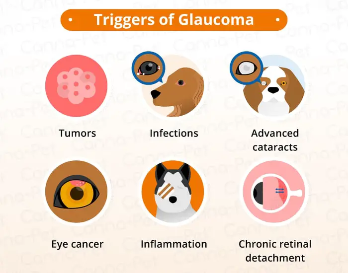
Types of Glaucoma: Secondary Glaucoma
Due to either iridocorneal angle blockage or impedance of aqueous drainage through the pupil, secondary glaucoma occurs as a result of other ocular diseases, including lens luxation, uveitis, neoplasia, intraocular hemorrhage, hyphaema, pigmentary, vitreous prolapse, intraocular surgery.
What are the Signs of Glaucoma in Dogs?
Some signs can occur very suddenly in acute narrow-angle glaucoma or secondary glaucoma. These significant signs are includes:
- Blindness in the affected eye.
- Ocular pain.
- Red-eye due to passive engorgement of the conjunctival and scleral vessels.
- Corneal edema.
- Dilated and non-responsive pupil due to paralysis of the iris sphincter muscle at high pressures.
In chronic glaucoma, all of the above signs may be seen with the addition of:
- Buphthalmos, an increase in the size of the eye.
- Tears in Descemet’s membrane, deep linear streaks seen in the cornea.
- Deep perilimbal corneal neovascularization.
- Iris atrophy, holes, or thinned areas of the iris stroma.
- Lens luxation.
- Cupping and atrophy of the optic nerve.
- Retinal vessel attenuation.
- Retinal atrophy.
What Dog Breeds are Prone to Glaucoma?
It is essential to know which breed is more likely to get glaucoma. The following breeds are associated with glaucoma-
- Toy poodle.
- Labrador Retriever.
- Samoyed.
- Beagle.
- Basset Hound.
- Boston Terrier.
- Cocker Spaniel.
- Shar-pei.
- Siberian Husky.
Diagnosis of Glaucoma in Dogs
The diagnostic procedure of glaucoma are as follows:
- Diagnosis of glaucoma depends upon the accurate identification of history- duration of the problem will affect potential to restore vision.
- Examination of the anterior and posterior segment of the eye for uveitis, lens luxation, and neoplasia.
- The appearance of the optic nerve and retina in chronic cases may predict the visual outcome.
- Tonometry ( IOP>25mmHg ).
- Schiotz pH applanation method.
- Gonioscopy (both eyes if possible, ‘normal’ eye if too much corneal edema in the glaucomatous eye)
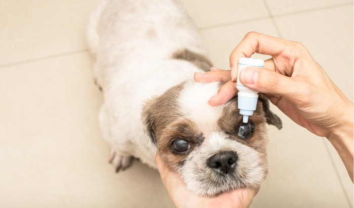
Treatment and Correction for Canine Glaucoma
Glaucoma is the outcome of many diseases in dogs’ eyes of the general system. The treatment and correction procedures are as follows:
- Mannitol and glycerol are usually prescribed your vets to reduce aqueous production by drawing fluid from the vitreous and aqueous humor into the surrounding vasculature.
- Dichlorphenamide or methazolamide maybe prescribe to control aqueous production by blocking carbonic anhydrase mediated buffering involved in aqueous secretion.
- The most commonly used drug, in this case, is Pilocarpine.
- Parasympathomimetics are believed to increase aqueous drainage by contracting the ciliary muscle and opening the trabecular meshwork.
- It is vital to reduce the IOP as soon as possible to control the risk of irreversible damage and blindness.
- Adrenaline or dipivefrine reduces IOP and also reduces aqueous production, and increases outflow.
- The ciliary body may be locally destroyed using either cryotherapy or laser therapy.
- Destruction of the ciliary process results in reduced aqueous production.
- Medical treatment must always be combined with surgical procedures in severe or advanced cases.
- Veterinary ophthalmologists use various surgical procedures to increase aqueous outflow.
- Ocular evisceration with prosthetic implants or enucleation.
- Pharmacological cycloablation is the injection of 25 mg.
- Gentamycin sulfate with 1 mg dexamethasone in a volume not to exceed 0.5 ml into the vitreous will destroy the ciliary process and reduces aqueous production.
Final Advice on Glaucoma in Dogs
Glaucoma is a common condition in aged dogs and few breeds of dogs. The condition is not life-threatening but irritating to dogs and its owner. If you identify the condition earlier as a dog owner, the condition may be corrected with minimum effort. In my article, I have discussed the most critical points on this condition. I think this article will help you a lot.