The horse skeletal system is one of the vital and most essential systems. The skeletal system forms the structure of the equine body, gives the body shape, is the storehouse of minerals, and protects vital organs of the body. A balanced skeletal system is essential for a sound horse. As a horse owner or veterinarian, knowledge of the equine skeletal system is of utmost importance.
Importance of Horse Skeletal System
The better proportion of the skeletal system ensures the horse’s best performance. A healthy and standard horse has 205 bones. Ligaments connect the bones, and the muscles are connected with the bones by tendons. The skeletal system of horses is divided into two major parts; axial skeleton and appendicular skeleton.
The function of the Skeletal System
The primary functions of skeletal systems are:
- The skeletal system gives the structure of the horse.
- They are the site for RBC production.
- Store macro minerals like calcium and phosphorus.
- Aids in locomotion.
- Protects vital organs like the brain, heart, and nervous system.
- Absorb concussion.
Equine Skeletal Anatomy: Types of Bones
The bones of the equine skeletal system have five primary types:
- Long Bone. The bones are mainly found in limbs which aid locomotion, produce red blood cells, and are a storehouse of minerals.
- Short Bones. The bones of the neck, hock, knee, and fetlock absorb the concussion.
- Flat Bones. The bones of the rib case, skull, scapula, and sternum protect the vital organs of the cranial, thoracic and abdominal cavities.
- Irregular Bones. The bones of the vertebral column protect the spinal cord.
- Sesamoid bones. The sesamoid bones are embedded ligaments which are also called navicular bones.
Horse Skeletal System: Axial Skeleton
The bones of axial skeletons are as follows:
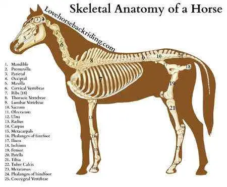
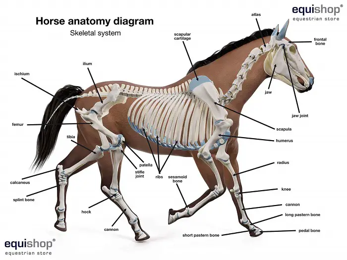
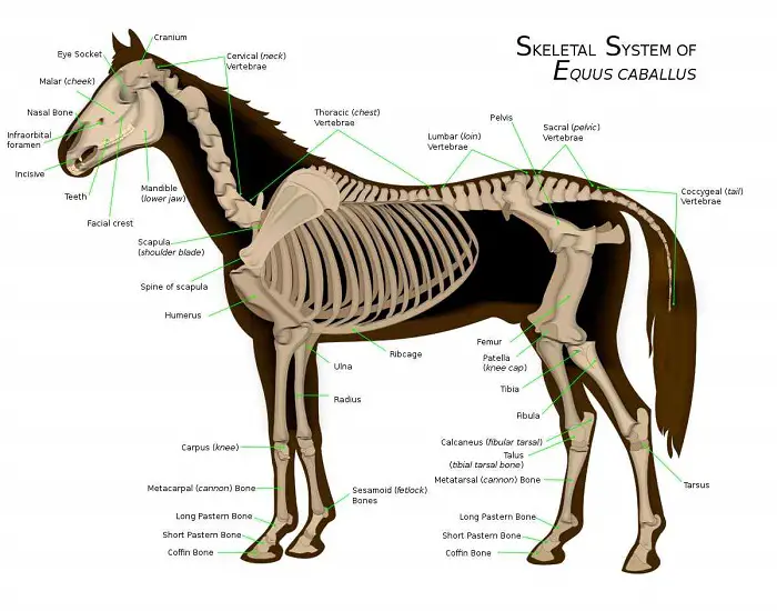
1. Skull. The skull of a horse has 34 bones and four cavities. The cavities are cranial, orbital, oral, and nasal. The cranial cavity protects the brain, the oral cavity hold and protects the eyes, the oral cavity passes the food particle, and the nasal cavity connects the respiratory system. The bones of the skull are as follows:
- Mandible.
- Maxillary bone.
- Nasal bone.
- Incisive bone.
- Lacrimal bone.
- Temporal bone.
- Frontal bone.
- Parietal bone.
- Occipital bone.
- Palatine bone.
- Zygomatic bone.
- Hyoid bone.
- Sphenoid bone.
- Vomer.
2. Ribcage. The ribcage is formed by 18 pairs of ribs and one sternum. The sternum has eight sternebrae that connect the eight pairs of true ribs. The ribcage protects the vital organs of the thoracic and abdominal cavities.
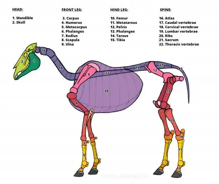
3. Vertebral Column. The vertebral column of a horse has a total of 54 bones. The bones are irregular and protect the spinal cord. The first cervical vertebrae are called the atlas, and the second cervical vertebrae are called the axis.
-
- Cervical vertebrae- 7.
- Thoracic vertebrae- 18.
- Lumbar Vertebrae- 5-6.
- Sacral vertebrae- 5.
- Caudal or coccigeal vertebrae- 18-23.
Equine Skeletal Anatomy: Appendicular Skeleton
The bones of the forelimbs and hindlimbs form the appendicular skeleton. The forelimbs do not directly connect to the vertebrae, as horses have no collar bones. The hindlimbs are connected to the axial skeleton at the pelvis. The forelimbs are connected to the body with muscles and tendons that give more flexibility for moving and jumping. The hindlimbs bear about 40% of the total body weight of a horse.
1. Bones of Fore Limb
The bones of the forelimbs are as follows:
- Scapula.
- Humorous.
- Radius.
- Ulna.
- Carpal bone (7-8).
- Metacarpal.
- Phalanges (first, second, and third).
2. Hind Limb/ Pelvic Limb
The bones of the hindlimbs of a horse are as follows:
- Pelvis (most prominent flat bone).
- Femur.
- Tibia.
- Fibula
- Tarsal bone (6 bones in two rows).
- Canon bone (third metatarsal bone)
- Splint bone (2nd and 4th metatarsal bone).
- Proximal sesamoid bone.
- Long pastern (first phalanx).
- Short pastern (second phalanx).
- Coffin bone (third phalanx)
- Navicular bones (distal sesamoid bone).
Horse Skeletal System: Joints of Horse
The joints are formed by attaching bones with ligaments. The joints have synovial cavities and fluid that help the movement of joints. The essential joints of horses are:
| Joints of Forelimb | Joints of Hindlimb |
| Shoulder | Hip |
| Elbow | Stifle |
| Knee | Hock |
| Fetlock | Fetlock |
| Pastern | Pastern |
| Coffin | Coffin |
Equine Skeletal Anatomy: Ligaments of Horses
The ligaments are the fibrous connective tissues that connect the bones. Ligaments give support and stabilize the skeleton. The ligaments have a limited blood supply. The major ligaments are:
- Nuchal ligament. Connect the dorsal surface of the cervical vertebrae.
- Supraspinous ligament. Connects the thoracic vertebrae after the nuchal ligament.
- Intercapital ligament. Connect first to the eleventh ribs.
- Suspensory ligament. Present back of the canon bone and divided into two branches.
- Interosseous ligament. Connect the canon bone with each splint bone.
- Proximal and Distal Check ligament.
- Planter ligament.
- Interosesamoidal ligament.
- Distal sesamoidal ligament.
- Imper ligament.
- Annular ligament.
- Sacrosciatic ligament.
Final Talk on Horse Skeletal System
A healthy and fit horse should have a balanced skeletal system. The skeletal system is the primary structure of the equine body. The knowledge of horse skeletal system will help you to keep your horse fit for work. You will be able to ensure the welfare of the horse.
My website has numerous articles on horse breeds, diseases, management, and general information. You can visit my website to learn more about this precious animal. You are welcome to drop your valuable comments.
