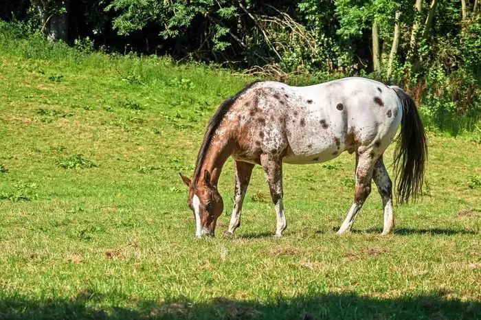
Leptospirosis in horses is a bacterial disease caused by several species of Leptospira spp. The organisms are found commonly throughout the world and are widespread in cattle herds. The bacteria can persist for many years in the soil where cattle have been housed. Leptospirosis in cattle causes abortion, stillbirths, decreased milk production, recurrent uveitis, and death in the cattle population. The disease is also found in the equine population.
Causes of Leptospirosis in Horses
Leptospirosis is divided into six significant serogroups, and these are then further divided into over 500 sero-varieties. There are seven significant sero-varieties found in horses. They are L autumnalis, L Bratislava, L canicola, L grippotyphosa, L hardjo, L icterohemorrhegica, and L Pomona.
Epidemiology of Equine Leptospirosis
Leptospirosis is very common in wild and domestic animals and can also produce diseases in humans. In particular areas, there will be one or more reservoir hosts that serve as reservoir hosts for the infection in particular areas. Leptospira will be transmitted to incidental hosts by shedding into the environment from a maintenance host. They can invade the mucous membrane, damaged skin, and migrate to various body organs of an incidental host.
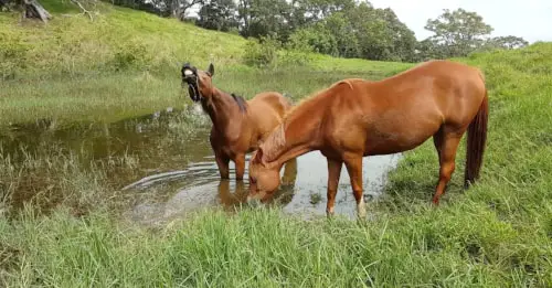
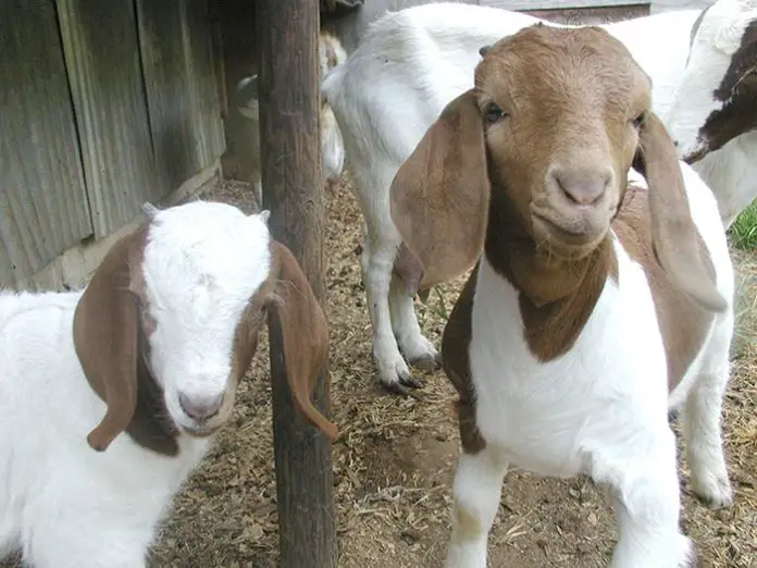
The contamination comes from contacting the infected animal’s urine through water, mud, bedding, or food. Cattle and swine can shed Leptospira in their urine for over a year, and horses may shed for few months. The maintenance cost can be wildlife such as deer, raccoons, and rodents.
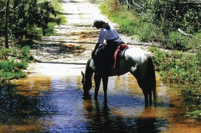
Leptospiral bacteria like warm and moist conditions and are found in many animals. Horses get the organism from drinking water that has been contaminated, or they may pick it up from pasture, hay, or grain infected by the urine of diseased animals. Horses can gain Leptospira from other horses, although most likely contracted it from diseased cattle, wild animals, and rodents.
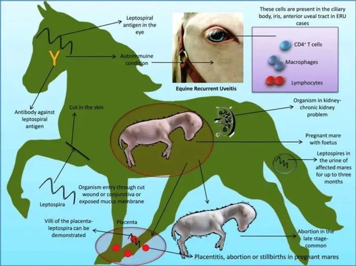
The frustrating thing about Equine leptospirosis and uveitis is that the uveitis may not occur for 18-24 months or more after the leptospirosis infection. In addition, while few animals are very ill with the disease, others may show no apparent signs of infection. Testing for leptospirosis at the time of uveitis may confirm that the disease was active simultaneously. Still, it is too late to take any action about the leptospirosis for that horse.
Clinical Signs of Leptospirosis in Horses
The clinical signs of leptospirosis include:
- Fever, anorexia, conjunctivitis, abnormal milk, and decreased milk production.
- Recurrent uveitis in horses was an immune-mediated reaction and a sequel to a Leptospira Pomona infection that occurred months or years previously.
- Acute signs include:
- varying degree of blepharospasm.
- lacrimation.
- photophobia.
- conjunctival and ciliary injection.
- Peripheral corneal edema and vascularization.
- Aqueous flare.
- Hypopyon or hyphema.
- Miosis.
- A swollen pale iris.
- Hypotonia.
- Vitreal inflammation.
- Retinal vasculitis or peripheral inflammation.
- No single case shows all these signs.
- Clinical signs often disappear, followed by reoccurrence.
- Severe infection can lead to calcific band keratopathy, permanent blindness, atrophy of the eye, and glaucoma.
- Pregnant mares can also have leptospirosis localize in the uterus or placenta and, subsequently, in the fetus, causing the death of the fetus.
- Equine abortions are commonly associated with L Pomona infection.
Diagnosis of Equine Leptospirosis
The standard diagnostic procedures of the disease are:
- The history makes the presumptive diagnosis of exposure of horses to cattle and wild animals.
- Specific clinical signs related to the reproductive system, eyes, and abortion inmates.
- Confirmatory diagnosis is made by testing of horse’s serum. A paired serum taken three weeks apart is essential for diagnosis. A fourfold change in titer to the organism in the paired sera would provide a presumptive diagnosis.
Treatment of Leptospirosis in Horses
The treatment of equine leptospirosis are:
- Penicillin and streptomycin is the primary drug of choice.
- Steroids are used topically or subconjunctivally to reduce the inflammation in uveitis.
- Atropine sulfate is used to reduce blepharospasm, lacrimation, and photophobia.
- Cyclosporin may be used during corneal ulceration.
Preventive Management of Leptospirosis in Horses
The preventive measures that are ensured by good management are:
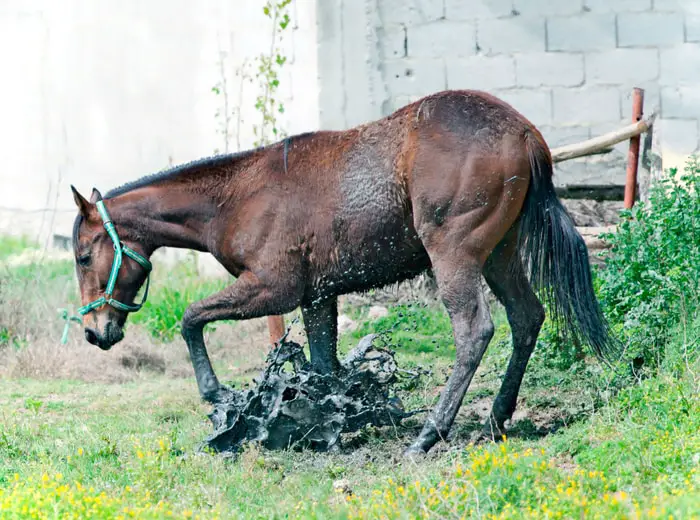
- Leptospira organisms can survive in water for 20 days and in manure for up to 61 days. It is therefore imperative to keep away animals from drinking stagnant water.
- Good management of manure.
- Good sanitation practices.
- Human hands can transmit infection from one animal to another and to even humans.
- Some precautions that can be taken include fencing water resources that wildlife can access.
- They are draining wet, muddy areas where horses are postured.
- Thoroughly disinfecting stalls and areas where animals have active leptospirosis.
- We are washing hands and clothes when in contact with sick animals.
- Vaccination of equine herds or individual animals in areas where leptospirosis is prevalent.