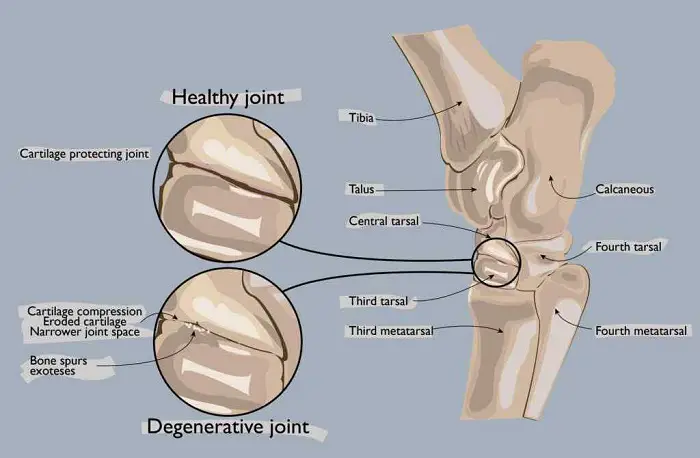
Bone spavin in horses is a common degenerative condition that affects the hock joint. The hock joint is located in the hind limb and is equivalent to the human ankle. Bone spavin typically affects the lower bones of the hock joint, particularly the tarsal bones.
Bone spavin is characterized by forming new bone and remodeling existing bone within the hock joint. Over time, this leads to bony growths, known as osteophytes, on the joint surfaces. These growths can cause inflammation, pain, and reduced range of motion in the affected joint.
Causes of Bone Spavin in Horses
The exact causes of equine bone spavin, also known as spavin, still need to be fully understood. However, several factors are believed to contribute to its development. These include:
- Conformation: Poor conformation, particularly in the hock region, can increase the risk of developing bone spavin. Horses with straight or sickle hocks may have abnormal stress distribution on the joint, leading to bony changes over time.
- Genetic Predisposition: Evidence suggests that certain breeds or bloodlines may be more susceptible to developing bone spavin. Genetics may play a role in determining the structural integrity of the hock joint and its ability to withstand stress and strain.
- Trauma or Injury: Direct trauma or repetitive stress to the hock joint, such as from poor shoeing, inappropriate exercise, or excessive strain during athletic activities, can contribute to the development of bone spavin. Injuries to the ligaments and other structures around the hock joint can disrupt its stability and lead to the formation of osteophytes.
- Joint Instability: Horses with unstable hock joints, either due to ligament laxity or congenital abnormalities, are more prone to developing bone spavin. Instability can result in abnormal joint movement and increased stress on the joint surfaces, leading to the formation of bony growths.
- Age and Wear and Tear: Bone spavin is more commonly observed in older horses, as the joint undergoes degenerative changes over time. The wear and tear associated with athletic activities and repetitive stress on the hock joint can contribute to the development of bony changes.
Contributing Factors of Equine Bone Spavin
In addition to the potential causes mentioned earlier, several contributing factors can increase the likelihood of a horse developing equine bone spavin:
- Overexertion and Intense Exercise: Horses engaged in intense athletic activities, such as racing, jumping, or strenuous training, are more susceptible to developing bone spavin. The repetitive stress and strain placed on the hock joint during these activities can accelerate joint degeneration and the formation of bony changes.
- Poor Hoof Balance and Shoeing: Incorrect hoof balance, improper trimming, or shoeing practices that do not adequately support the hock joint can contribute to the development of bone spavin. These factors can result in abnormal loading patterns on the joint and increased stress, leading to joint damage over time.
- Obesity and Overweight: Excessive body weight stresses the hock joint. Overweight horses have a higher risk of developing joint problems, including bone spavin. Maintaining a healthy body condition through proper diet and exercise can help reduce the risk.
- Nutritional Imbalances: Poor nutrition, particularly a diet lacking essential minerals and vitamins necessary for joint health, can contribute to developing joint disorders, including bone spavin. A balanced diet and appropriate supplements can help support joint health and reduce the risk of degenerative conditions.
- Previous Joint Injuries: Horses that have experienced prior hock joint injuries, such as sprains or fractures, may be more prone to developing bone spavin. The trauma and damage to the joint structures can disrupt the standard joint mechanics, leading to osteoarthritis and bony changes.
- Aging and Degenerative Changes: As horses age, the joint tissues naturally undergo degenerative changes. The cartilage within the hock joint may deteriorate over time, increasing the risk of bone spavin development. Age-related factors can make the joint more susceptible to the effects of other contributing factors.
Clinical Signs of Degenerative Joint Disease in Horses
Equine bone spavin, also known as spavin, can cause various clinical signs in affected horses. The severity and specific signs can vary depending on the stage and extent of the condition. Here are some common clinical signs associated with equine bone spavin:
- Lameness: Lameness is one of the primary signs of bone spavin. A gradual onset often characterizes the lameness. It may be more apparent during certain activities, such as turning, backing up, or starting to move after rest. The severity of lameness can vary, ranging from mild to severe, and may worsen with exercise or prolonged activity.
- Stiffness: Horses with bone spavin often exhibit stiffness, particularly in the hock joint. They may have difficulty flexing or extending the joint fully, leading to a shortened stride or altered gait. The stiffness is usually more pronounced when the horse is first moved after rest and may improve with continued exercise.
- Swelling and Heat: Inflammation within the hock joint can cause localized swelling and increased warmth over the affected area. The swelling may be mild to moderate and can sometimes be palpable. However, in some cases, the swelling may not be visibly apparent.
- Reduced Performance: Horses with bone spavin may experience a decline in performance. They may struggle with tasks that require flexion or extension of the hock joint, such as jumping, collection, or speed work. A decrease in the horse’s ability to engage the hind end or reluctance to perform specific movements may be observed.
- Changes in Behavior: Some horses with bone spavin may exhibit changes in behavior or attitude. They may become irritable, resistant, or unwilling to perform specific movements or tasks that cause discomfort or pain. The horse may show discomfort during grooming or touching the affected area.
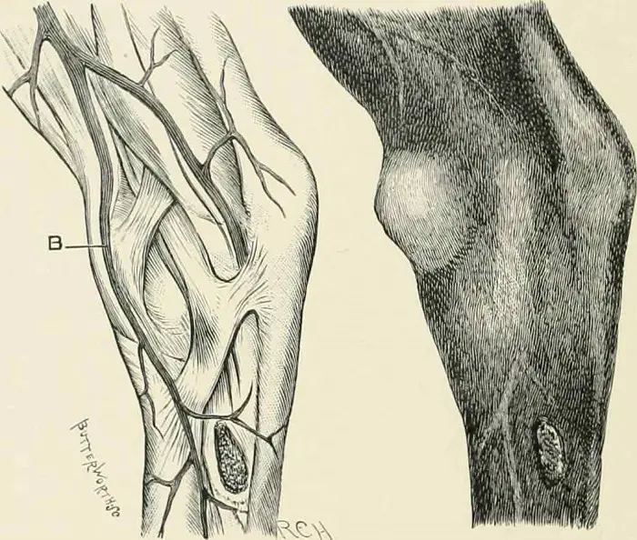
- Joint Enlargement: As bone spavin progresses, the hock joint may develop bony growths or osteophytes. These growths can result in joint enlargement, particularly around the lower part of the hock. The size and visibility of the enlargement can vary depending on the severity of the condition.
Diagnosis of Bone Spavin
The diagnosis of bone spavin in horses typically involves a combination of clinical evaluation, lameness assessment, and diagnostic imaging. Here is an overview of the standard methods used for diagnosing bone spavin:
- Physical Examination: The veterinarian will thoroughly examine the horse, paying particular attention to the hock region. They will assess the horse’s gait, observe for lameness or stiffness, and palpate the hock joint to check for swelling, heat, and pain response.
- Lameness Evaluation: The horse will undergo a lameness assessment to evaluate the severity and characteristics of lameness. This may involve observing the horse’s movement on different surfaces, lunging, or performing flexion tests to provoke lameness and localize the affected area.
- Diagnostic Imaging: Radiography (X-rays) is the most common imaging modality to diagnose bone spavin. X-rays allow visualization of the bony structures and can reveal the presence of osteophytes, joint changes, and other abnormalities. The hock joint is typically imaged from various angles for a comprehensive assessment.
- Additional Imaging: In some cases, additional imaging techniques may be used to evaluate the hock joint further. This can include ultrasound or nuclear scintigraphy (bone scan). Ultrasound can provide detailed images of soft tissues, while a bone scan can help identify areas of increased bone activity and inflammation.
- Diagnostic Analgesia: Diagnostic analgesia, commonly called a “joint block,” may be performed to confirm the source of lameness and its specific location. Local anesthetics are injected into specific areas around the hock joint, and lameness improvement afterward can help pinpoint the affected structures.
- Joint Fluid Analysis: In some instances, joint fluid analysis may be performed. A sample of synovial fluid can be obtained via joint aspiration and analyzed for signs of inflammation, infection, or other abnormalities.
The combination of these diagnostic methods allows veterinarians to assess the presence and severity of bone spavin, identify any other concurrent conditions, and rule out alternative causes of lameness.
Differential Diagnosis of Bone Spavin in Horses
When evaluating a horse with suspected bone spavin, veterinarians must consider other conditions that can cause similar clinical signs. The following are some of the differential diagnoses that should be considered:
- Osteoarthritis: Osteoarthritis can affect various joints in horses, including the hock joint. It shares many similarities with bone spavin regarding clinical signs and joint changes. Diagnostic imaging and joint fluid analysis may help differentiate between the two conditions.
- Bog Spavin: Bog spavin is characterized by swelling and distension of the tibiotarsal (hock) joint capsule due to excessive joint fluid. It can cause lameness and hock stiffness, mimicking some signs of bone spavin. Radiographs and joint fluid analysis can aid in distinguishing between the two conditions.
- Hock Injuries: Traumatic injuries to the hock, such as fractures, ligament tears, or soft tissue injuries, can result in lameness and joint swelling. An accurate history, lameness evaluation, and diagnostic imaging can help identify specific injuries and differentiate them from bone spavin.
- Osteochondrosis (OCD): Osteochondrosis is a developmental joint disease that can affect the hock joint in young horses. It involves abnormal cartilage development, leading to loose fragments within the joint. OCD can cause lameness and hock joint swelling, similar to bone spavin. Radiographs or advanced imaging techniques can help identify OCD lesions.
- Suspensory Ligament Injuries: Injuries or inflammation of the suspensory ligament can cause lameness and hock stiffness. The pain associated with suspensory ligament injuries can sometimes be mistaken for bone spavin. Ultrasonography and diagnostic anesthesia can assist in diagnosing suspensory ligament injuries.
- Infectious Arthritis: Bacterial or fungal joint infections can cause severe lameness, joint swelling, and systemic signs of illness. Infectious arthritis should be considered, especially in sudden-onset severe lameness or rapidly progressing joint swelling. Joint fluid analysis and culture can help diagnose infectious arthritis.
- Other Causes of Lameness: Lameness in horses can also be caused by issues such as hoof abscesses, tendon/ligament injuries, hoof balance problems, navicular syndrome, or other joint diseases. A thorough examination, including diagnostic imaging, is crucial to identify the underlying cause of lameness accurately.
What is the Best Treatment for Bone Spavin in Horses?
The treatment for bone spavin in horses aims to manage the symptoms, reduce pain, and slow down the condition’s progression. The specific treatment plan may vary depending on the severity of the disease, the horse’s response, and the intended use of the horse. Here are some standard treatment options for bone spavin:
- Rest and Controlled Exercise: Resting the horse can help reduce inflammation and allow the joint to heal. After the rest period, controlled exercise can help improve joint mobility and promote the strengthening of supportive structures. It’s essential to consult a veterinarian or equine rehabilitation specialist to develop an appropriate exercise program for the horse’s needs.
- Non-Steroidal Anti-Inflammatory Drugs (NSAIDs): NSAIDs, such as phenylbutazone (bute) or flunixin meglumine, can help alleviate pain, reduce inflammation, and improve the horse’s comfort. These medications are commonly used in the short term or during flare-ups to manage pain associated with bone spavin. However, a veterinarian should monitor and guide long-term use to minimize potential side effects.
- Joint Supplements: Various oral supplements contain glucosamine, chondroitin sulfate, hyaluronic acid, and omega-3 fatty acids. These supplements support joint health, promote cartilage integrity, and reduce inflammation. While scientific evidence for their effectiveness is mixed, some horse owners and veterinarians report positive results.
- Intra-articular Medications: In cases where pain and inflammation persist despite other treatments, intra-articular injections may be considered. Corticosteroids or other medications can be injected directly into the affected joint to reduce inflammation and relieve pain. A veterinarian typically performs these injections, which should be used judiciously due to potential risks and side effects.
- Physical Therapy and Rehabilitation: Physical therapy modalities, such as therapeutic ultrasound, laser, or electromagnetic therapy, may help manage pain, reduce inflammation, and promote healing. Controlled exercise programs, including exercises for joint mobilization and strengthening, can also be beneficial under the guidance of a qualified equine rehabilitation specialist.
- Corrective Shoeing and Hoof Balance: Proper shoeing and hoof balance can help alleviate stress on the hock joint and provide support. Working with an experienced farrier or veterinarian knowledgeable in corrective shoeing can be beneficial in optimizing the horse’s comfort and biomechanics.
- Surgical Interventions: In severe cases of bone spavin that do not respond to conservative treatments, surgical options may be considered. These can include arthroscopic procedures to remove or smooth out bony growths or surgical joint fusion to stabilize and alleviate pain. These procedures are typically reserved for cases where other treatments have failed or the horse’s quality of life is significantly affected.
Prevention of Degenerative Joint Disease
While some factors contributing to bone spavin, such as conformation and genetic predisposition, cannot be fully controlled, specific measures can help reduce the risk or delay the onset of bone spavin in horses. Here are some prevention strategies:
- Proper Nutrition: Providing a well-balanced diet that meets the horse’s nutritional requirements is crucial for overall joint health. Ensuring the horse receives appropriate levels of vitamins, minerals (including calcium and phosphorus), and essential fatty acids can support the integrity of joint tissues.
- Regular Exercise and Conditioning: Regular exercise and appropriate conditioning can help maintain joint health and strengthen supporting structures. Increasing the horse’s workload and incorporating varied exercises can help build muscle strength and improve overall joint function.
- Maintaining Optimal Body Condition: Avoiding obesity and maintaining an appropriate body condition score (BCS) is essential to reduce excessive stress on joints, including the hock joint. Proper nutrition, exercise, and monitoring of the horse’s body condition can help prevent excessive weight gain.
- Regular Veterinary Care: Routine veterinary examinations and preventive healthcare measures are essential for detecting early joint problems or predisposing factors. Regular check-ups, dental care, deworming, and vaccinations can contribute to the overall health and well-being of the horse.
- Proper Hoof Care and Shoeing: Maintaining good hoof balance and appropriate shoeing practices can help promote proper weight distribution and reduce stress on the hock joint. Working with a skilled farrier who understands the horse’s conformation and individual needs can help optimize hoof health and minimize biomechanical issues.
- Environmental and Management Factors: Providing horses with suitable turnout conditions, including adequate space for movement, proper footing, and appropriate shelter, can contribute to joint health. Minimizing excessive and repetitive stress on joints through proper training techniques, avoiding sudden high-intensity exercise, and providing sufficient rest periods can also help prevent joint problems.
- Monitoring and Early Intervention: Regularly monitoring the horse’s gait, movement, and behavior can help detect signs of lameness or joint discomfort at an early stage. Prompt veterinary evaluation and intervention can help address any issues and prevent the progression of joint problems.
Final Words on Equine Bone Spavin
Equine bone spavin is a degenerative condition affecting horses’ hock joints. The formation of bony growths characterizes it and can cause lameness, stiffness, and reduced performance. While the exact causes are not fully understood, factors such as conformation, genetics, trauma, and intense exercise can contribute to its development.
Diagnosing bone spavin involves a thorough veterinary examination, including lameness evaluation and diagnostic imaging. Differential diagnoses should be considered to rule out other conditions with similar symptoms. Treatment aims to manage pain, reduce inflammation, and slow disease progression. It may include rest, controlled exercise, medications, joint supplements, physical therapy, and, in severe cases, surgery.
Prevention strategies for bone spavin include proper nutrition, exercise, hoof care, and regular veterinary care. While prevention cannot be guaranteed, these measures can help reduce the risk and maintain joint health.
Horse owners must work closely with their veterinarians to develop individualized treatment and management plans. Regular monitoring and proactive care are essential for the well-being and soundness of horses affected by bone spavin. Owners can optimize the horse’s comfort and quality of life by staying attentive to the horse’s needs, providing appropriate care, and seeking veterinary guidance.