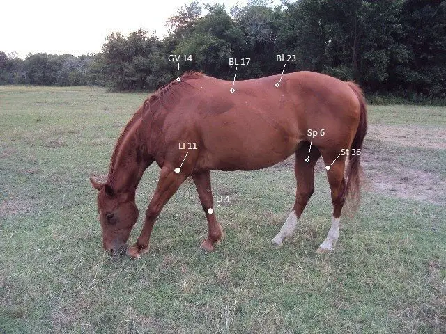
Pigeon fever in Horses is the most common term of the bacterial infection of horses caused by Corynebacterium pseudotuberculosis var equi. Another name of the disease is Dry land Distemper. The C pseudotuberculosis is a localized bacteria and causes an abscess in the lower abdomen and pectoral region of horses. The name Pigeon Fever given as the horse’s chest becomes the “Pigeon breast” like appearance due to the swelling of the abscess. The disease is most common in California. Other ruminants like sheep, goat, and cattle are also affected by bacteria with similar clinical signs. The cross-species transmission of the disease occurs between horses and cattle.
What is pigeon fever caused by?
Corynebacterium pseudotuberculosis causes a syndrome of ulcerative lymphangitis or abscess formation. Inflammation of hair follicles and abortion have also been reported. The bacterium is a Gram-positive, facultative anaerobic and non-motile that survives well in the environment and can infect other species, including man.
Epidemiology of Pigeon Fever in Horses
The disease occurs throughout the world in areas where horses are kept. However, there is a marked variation in clinical signs in geographic locations of the world. Ventral, mid-line, pectoral, abdominal and inguinal abscesses are frequent presentations in areas of the western United States, whereas ulcerative lymphangitis is more typical elsewhere. The rate of occurrence of the disease varies from year to year, and outbreaks occur. Common names of the abscess form of the disease are Corynebacterium pseudotuberculosis,dry-land distemper, Colorado distemper, or pigeon breast.
Folliculitis (contagious pustular dermatitis, “contagious acne,” or “Canadian horse-pox”) caused by C pseudotuberculosis results in pustules that are sometimes distributed in areas where tack or harness make contact with the skin. The disease may be spread by contaminated grooming utensils. Trauma to the skin, existing folliculitis, or seborrhoea may predispose the horse to infection. Ulcerative lymphangitis is a less uncommon disease that is usually associated with poor sanitation. Environmental contamination may be done by open draining tracts.
Pathogenesis of Pigeon Breast in Horses
The organism finds a portal of entry through damaged mucous membranes or lacerated skin surfaces such as may be caused by badly fitting tack. Biting inserts may also provide a port of entry. This phenomenon may explain the seasonal occurrence of the disease, particularly for the abscess form.
The disease may establish itself locally or spread via blood or lymph. Entry into the lymphatic system of the limb is likely to be through local skin lesions and causes in typical ulcerative lymphangitis.
The incubation period for the infection can be long and variable from weeks to months. The organism produces exotoxin is thought to be significant in abscess formation.
What are the Symptoms of Pigeon Fever in Horses?
- Typical abscess formation occurs mostly in the pectoral region and occasionally in the abdomen along the inguinal region and ventral midline.
- The abscess is often thick-walled and can progress in size. Ventral and inguinal edema can be seen in association with abscesses.
- Fever, depression, and anorexia may be present but are atypical. Axillary form abscesses may result in gait dysfunction before abscesses are identified.
- Internal abscesses may cause signs of weight loss or abnormal pain.
- The lower limb from the disease may be initially identified as a non-descript swelling that can produce severe lameness.
- Nodules of subcutis frequently develop, often in the area of the pastern joint, but they can spread to other sites.
- Nodules may range from a few millimeters to severe cm in diameter.
- Ruptured nodules discharge creamy pus and result in an ulcer of the skin surface at the site of rupture.
- Lymphatics draining the area may become firm and enlarged.
- Nodules and ulcers can develop along these lymphatics. Repetition of the disease may occur after resolution.
- The pustules and papules of the skin form of the disease usually occur in groups.
- These are often associated with frequently damaged skin (from harness and thick) but can be more diffuse.
- Smaller papules may develop into pustules.
- The ruptures of pustules lead to a crust formation.
- Abscess are usually not pruritic but may be painful.
Clinical Pathology and Necropsy of Dry-Land Distemper
Elevations in white blood cell count for fibrinogen may be identified early in the course of the disease. Chronically diseased animals may be haematologically normal. Thick-walled abscesses are typical post-mortem findings. Abscesses usually contain a tan-colored inflammatory fluid, whereas pustules may contain greenish-white pus. Abscesses are identified along with the lymphatic system in peripheral limbs.
Diagnosis of Pigeon Fever in Horses
- Characteristic clinical signs such as ulcerative lymphangitis, large abscesses in the pectoral region, or cellulitis with multiple small skin erosion of one or more limbs may suggest a C pseudotuberculosis infection.
- A definitive diagnosis requires a bacterial culture.
- Pure cultures can be obtained from the lesions.
- Abdominocentesis may identify abdominal abscesses, but the normal peritoneal liquid does not rule out their presence be obtained.
- Ultrasonography can be useful in identifying internal abscesses in inguinal abdominal or axillary regions.
Treatment of Dry-Land Distemper
The surgical section into open drainage of abscesses is often required and aids the resolution of infection. Pectoral and inguinal abscesses are often very deep and may require a deep incision or the use of large trocar drains. Ultrasonographic localization of abscesses can be beneficial. Daily flushing and local therapy may be required to maintain open drainage. Anti-inflammatory therapy may be useful.
The efficacy of antibiotic therapy is doubtful. The in vitro susceptibility of C pseudotuberculosis is wide. Procaine penicillin (20000 IU/kg IM b.i.d.), potassium penicillin (20000-40000 IU/kg IV q.i.d.), and sulphatrimethoprim (5 mg/kg of the trimethoprim portion twice) are commonly used. The failure of antibiotic treatment may be a result of the intracellular nature of the organism or the inability of antibiotics to reach optimum levels in large, thick-walled abscesses.
How do you prevent pigeon fever in horses?
- Improvement of sanitation and a restriction in constant exposure to moisture is imperative in reducing the incidence of the peripheral limb from the disease.
- Preventing skin abrasion from tack or harness is helpful. Culling or quarantine of infected horses reduces environmental spread.
- Pigeon fever is susceptible to most disinfectants.
- Fly control and the use of screened stables may decrease insect vector spread.
- Claims have been made for the successful application of autogenous bacterins, that have been used on a limited scale, without serious adverse effects.